Macroscopic and Microscopic Findings of Microsporum gypseum
Abstract
Keywords
Macroconidium Microsporum gypseum Morphology
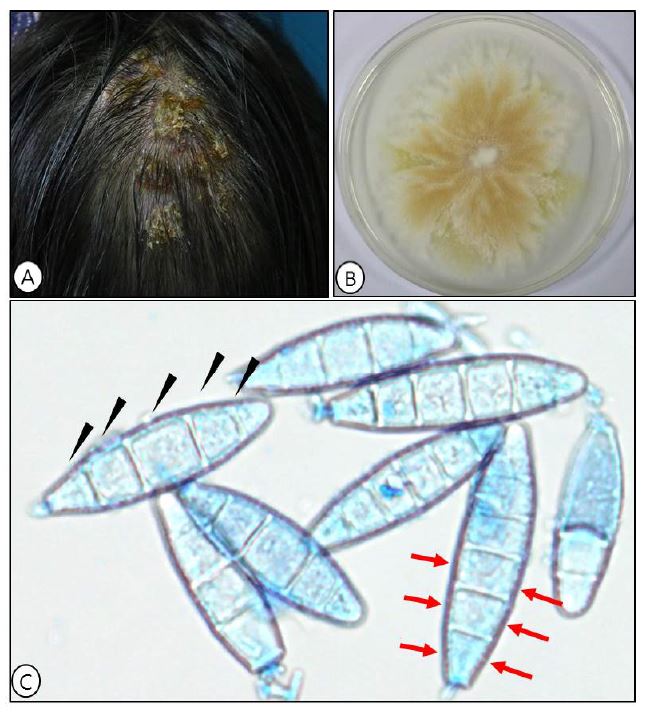
Microsporum (M.) gypseum is a geophilic dermatophyte that is prevalently distributed world-wide[1]. Particular transmissions occur in warm, humid, and rural areas among farmers and children. Clinical forms may present scaly patches of variable shapes and colors on the skin[2]. Tinea capitis is the typical clinical form and pus frequently occurs from palpitated hair follicles[1]-[2].
Macroscopic morphology of M. gypseum is characterized by powdery to granular texture with a color range of white to yellowish, often termed cinnamon colored (
Fig. 1A). The powdery appearance is imparted by heavy sporulation on the older mycelium and the edges of the colony can exhibit pleomorphism described as scalloped to ragged nature
[3]. The reverse shows no specific pattern indicative of the species. In microscopic morphology, short, spindle-shaped macroconidia are borne directly on hyphae. Characteristic macroconidium is divided in to 4 to 6 septated cells with thick walls and smooth borders (
Fig. 1B). Unlike M. canis, macroconidia are more numerous and are less barrel-shaped with fewer cells
[4]. Microscopic difference may help distinguish from other dermatophytes, however, culture and genetic studies should discern accurate diagnoses
[5].
References
1. Silva KVS, Lima MIO, Cardoso GN, Santos AS, Silva GS, Pereira FO. Inibitory effects of linalool on fungal pathogenicity of clinical isolates of Micro- sporum canis and Microsporum gypseum. Mycoses 2017;60:387-393
Crossref
Google Scholar
2. Romano C, Asta F, Massai L. Tinea incognito due to Microsporum gypseum in three children. Pediatr Dermatol 2000;17:41-44
Crossref
Google Scholar
3. Mihali CV, Buruiana A, Turcus V, Covaci A, Ardelean A. Morphological aspects of fruiting bodies in Microsporum gypseum on Sabouraud's dextrose agar medium. Ann of RSCB 2011;16:85-92
Google Scholar
4. Demange C, Contet-Audonneau N, Kombila M, Miegeville M, Berthonneau M, De Vroey C, et al. Microsporum gypseum complex in man and animals. J Med Vet Mycol 1992;30:301-308
Google Scholar
5. Sharma R, Gupta S, Asati DP, Karuna T, Purwar S, Biswas D. A pilot study for the evaluation of PCR as a diagnostic tool in patients with suspected der- matophytoses. Indian Dermatol Online J 2017;8:176 -180
KJMM
2017 September;22(3):144-145(2). http://dx.doi.org/10.17966/KJMM.2017.22.3.144 Epub 2017 September 29
Copyright © 2017 by Korean Journal of Medical Mycology
Language
Korean/English
Author
Osung Kwon; Department of Dermatology, School of Medicine, Catholic University of Deagu, Daegu, Korea
Joonsoo Park; Department of Dermatology, School of Medicine, Catholic University of Deagu, Daegu, Korea
Yong Joon Bang; Catholic Skin Clinic, Medical Mycology, Daegu, Korea
Hyun Chung; Department of Dermatology, School of Medicine, Catholic University of Deagu, Daegu, Korea
Corresponding
Hyun Chung, Department of Dermatology, School of Medicine, Catholic University of Daegu, 33 Duryugongwon-ro 17gil, Namgu, Daegu, 42472, Korea. Tel: +82-53-650-4161, Fax: +82-53-650-4891, e-mail: hyunch@cu.ac.kr
Publication history
Received 30 July 2017;
Revised 7 September 2017;
Accepted 16 September 2017.
Acknowledgements
This is an Open Access article distributed under the terms of the Creative Commons Attribution Non-Commercial License (http://creativecommons.org/licenses/by-nc/3.0/) which permits unrestricted non-commercial use, distribution, and reproduction in any medium, provided the original work is properly cited.
Osung Kwon
Department of Dermatology, School of Medicine, Catholic University of Deagu, Daegu, Korea
Joonsoo Park
Department of Dermatology, School of Medicine, Catholic University of Deagu, Daegu, Korea
Yong Joon Bang
Catholic Skin Clinic, Medical Mycology, Daegu, Korea
Hyun Chung
Department of Dermatology, School of Medicine, Catholic University of Deagu, Daegu, Korea
- Download : 804
- Image : 0
- DOC : 0
- XLS : 0
- PDF : 804
Since epub date 2017 September 29