pISSN : 3058-423X eISSN: 3058-4302
Open Access, Peer-reviewed
pISSN : 3058-423X eISSN: 3058-4302
Open Access, Peer-reviewed
Osung Kwon,Joonsoo Park,Hyungrok Kim,Jae Bok Park
http://dx.doi.org/10.17966/KJMM.2016.21.4.138 Epub 2016 December 27
Abstract
Keywords
Malassezia Seborrheic dermaitits Spore
Seborrheic dermatitis is a chronic inflammatory disease characterized by yellowish adherent scales in sebaceous gland rich regions such as scalp and face[1]. Although the pathogenesis encompasses genetic and environmental factors, numerous studies reveal Malassezia colonization on the scalp and a positive correlation of Malassezia with the severity of seborrheic dermatitis[1],[2]. Clinical response to antifungal agents such as ketoconazole and ciclopirox has contributed a crucial role of Malassezia in the pathogenesis of seborrheic dermatitis1.
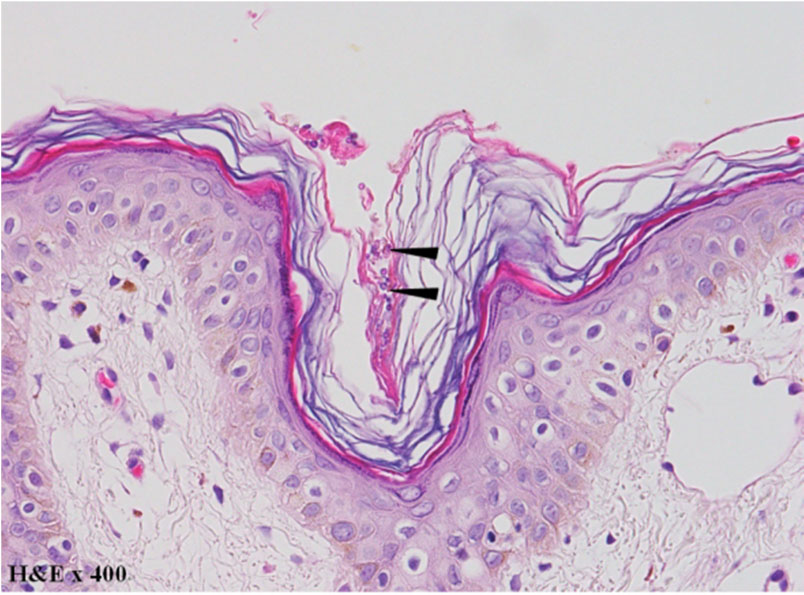
Histopathogy of seborrheic dermatitis with Malassezia infection reveals hyperkeratotic stratum corneum with numerous hyphae and spores often referred as spaghetti and meatball in light microscopy. Morphologically, the spores are round to ovoid in shape, approximately 1.5~4.5 μm in diameter depending on the species and are stained basophilic under hematoxylin and eosin staining (Fig. 1). While the spores are embedded within the keratin layer of the epidermis, some species may involve the follicles and the surrounding dermis. Due to various etiologic factors of seborrheic dermatitis, Malassezia infection should always be ruled out by identifying spores in a typical presentation of the disease.
Conflict of interest
In relation to this article, I declare that there is no conflict of interest.
References
1. Borda LJ, Wikramanayake TC. Seborrheic dermatitis and dandruff: a comprehensive review. J Clin Investig Dermatol 2015;3:10.13188/2373-1044.1000019
Crossref
Google Scholar
2. Kim SY, Kim SH, Kim SN, Kim AR, Kim MJ, Park WS, et al. Isolation and identification of Malassezia species from Chinese and Korean patients with seborrheic dermatitis and in vitro studies on their bioactivity on sebaceous lipids and IL-8 production. Mycoses 2016;59:274-280
Crossref
Google Scholar
