pISSN : 3058-423X eISSN: 3058-4302
Open Access, Peer-reviewed
pISSN : 3058-423X eISSN: 3058-4302
Open Access, Peer-reviewed
Ji Yeon Hong,Joon Seok,Won Jong Oh,Nam Ju Moon,Kapsok Li,Seong Jun Seo
http://dx.doi.org/10.17966/KJMM.2016.21.2.34 Epub 2016 July 03
Abstract
Protothecosis is an unusual human infection, caused by the genus prototheca, especially Prototheca wickerhamii. A 80-year-old immunocompetent man presented with a 1-month history of multiple reddish brown lobulated plaques on the scalp and face. He denied any history of trauma and had no evidence of underlying diseases such as diabetes mellitus or malignancy. On histopathological examination, characteristic morula-like sporangia in the dermis was revealed. After treatment with oral itraconazole for 8 weeks, the patient's skin lesions are almost healed.
Keywords
Cutaneous protothecosis Prototheca
서 론
프로토테카증은 단세포의 호기성, 비엽록소성 조류인 Prototheca 균에 의해 유발되는 드문 기회 감염증으로, 인체에 발생하는 경우 주로 Prototheca wickerhamii가 원인이 된다[1]. 피부 프로토테카증의 경우 인체 면역결핍바이러스 감염, 후천성면역결핍증, 당뇨, 전신 스테로이드 장기 복용 등 의 위험 요인을 가진 면역저하자에서 발생하는 경우가 많으나 정상 면역자에서도 드물게 발생할 수 있다[2]. 정상 면역 환자에서 발생하는 경우 주로 국한된 병변의 구진과 농포 형태를 보인다[3]. 호발 부위는 얼굴과 사지의 노출부로, 외상이 선행된 부위에 기회 감염되는 것이 주된 감염 경로이다[4].
저자들은 정상 면역을 가진 80세 남자 환자에서 외상력 등 특이 병력 없이 발생한 두피와 안면부의 다발성 홍반성 판의 임상 양상을 보인 피부 프로토테카증 1예를 경험하고 문헌 고찰과 함께 보고한다.
증 례
환 자: 이OO, 80세, 남자
주 소: 두피와 미간 상안검, 입 주위에 발생한 다양한 크기와 모양의 다발성 홍반성 판
현병력: 내원 1개월 전 두피에 작은 홍반성 판이 처음 발생하였으며 소양감 동반되어 타 병원에서 타크로리무스0.03% 연고만 처방 받아 도포하였으나 점차 얼굴로 번지면서 안면부 전반에걸쳐 다양한 크기의 다발성 홍반성 판으로 진행되어 내원함.
과거력 및 가족력: 수년 전부터 고혈압 진단받아 치료 중이며 집 앞 마당을 가꾸는 일을 종종 하였으며 외상력 없고 그 외 특이 과거력 및 가족력 없음.
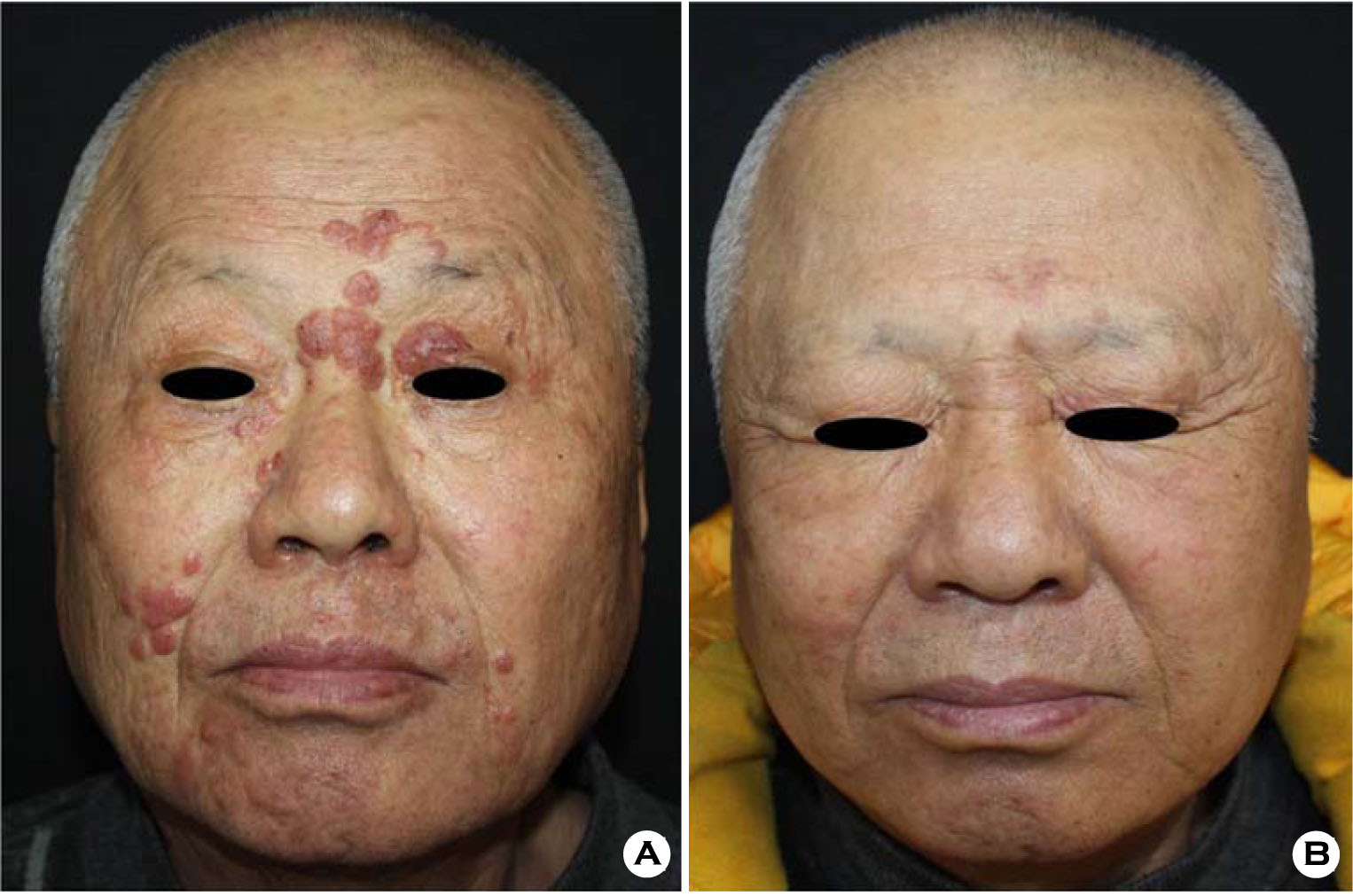
피부 소견: 두피와 미간, 상안검, 입 주위에 발생한 다양한 크기와 모양의 다발성 홍반성 판 (Fig. 1A).
이학적 소견: 피부 소견 외에 특이 사항 없음.
검사 소견: 일반혈액 검사상 WBC 12,770/μl로 증가되었으며 BUN/Cr이 23/1.27로 약간 상승한 소견 이외 간기능 검사를 포함한 다른 혈액 검사, 소변 검사 상 특이 소견은 없었다.
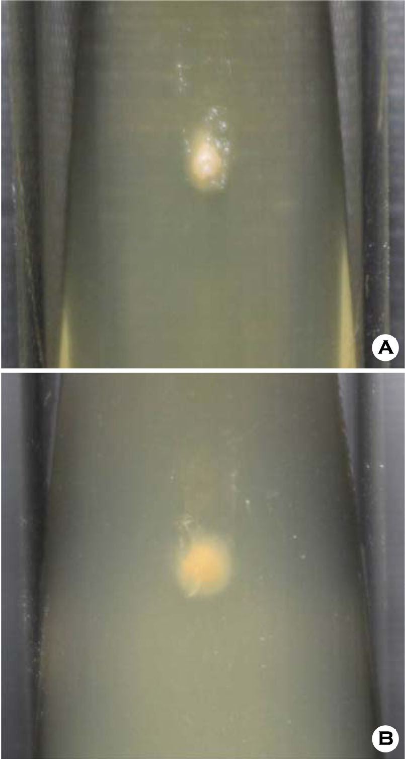
진균학적 소견: 펀치로 절제한 조직을 Sabouraud dextrose agar 사면 배지에 심어 실온에서 2주간 배양 후 백색 크림 형태의 집락이 형성되었다 (Fig. 2).
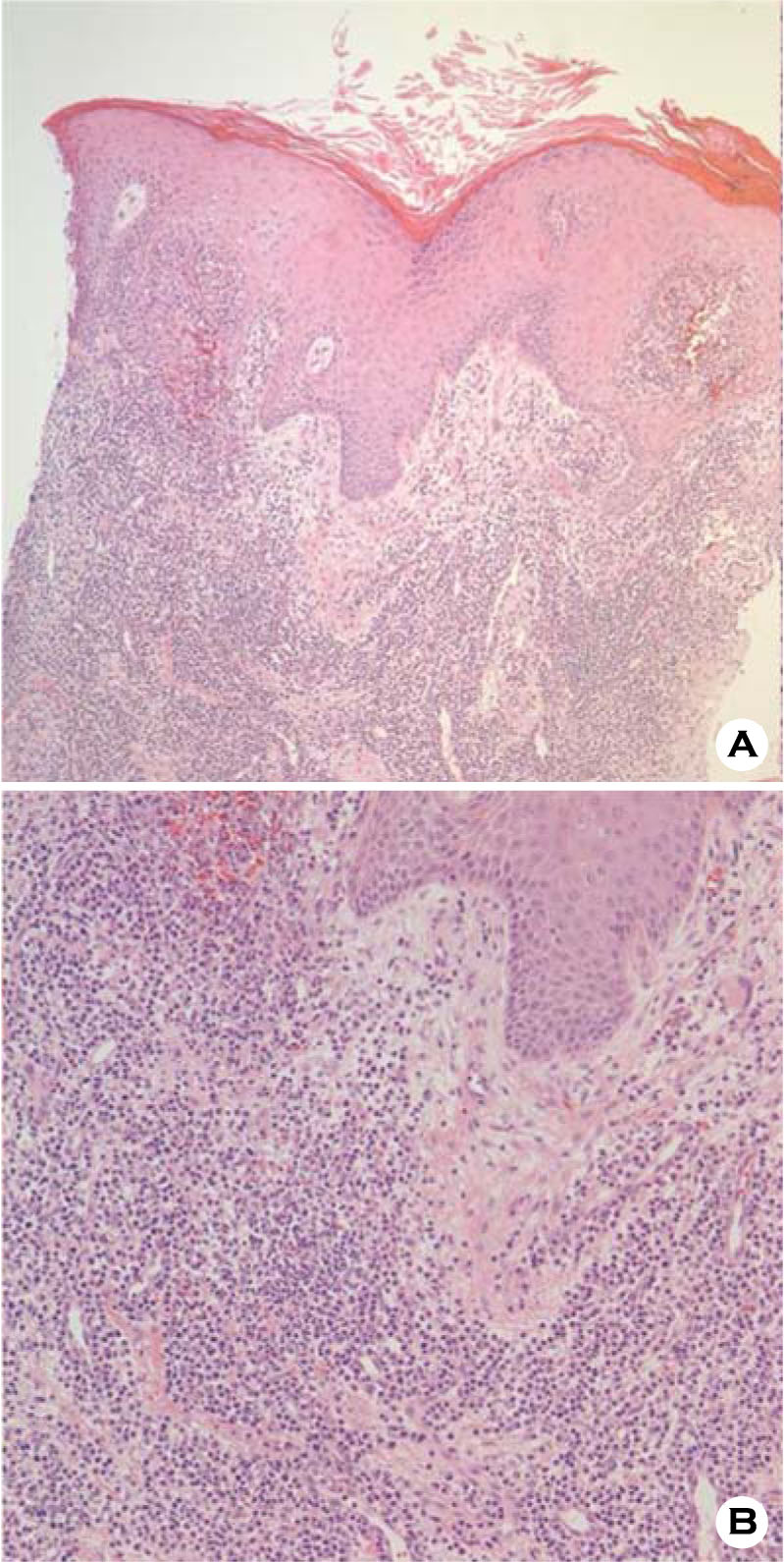
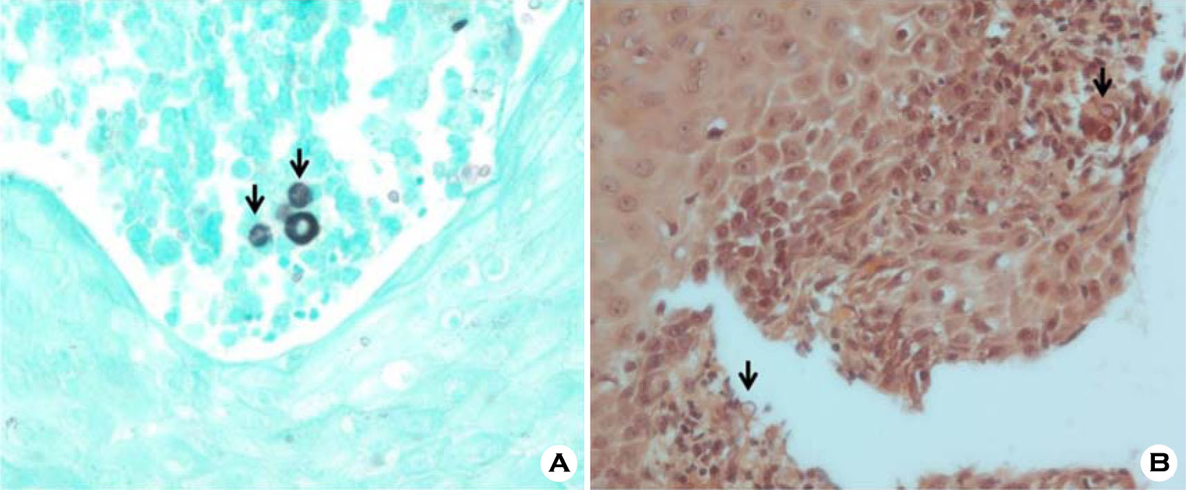
병리 조직학적 소견: 판의 중심부에서 시행한 조직 검사 상, H&E 염색에서 표피의 과각화증, 가시세포증이 관찰되었고, 진피 전반에 걸쳐 거대 세포들과 함께 다수의 림프구, 형질세포와 일부 호산구가 혼재된 소견을 보였으며 둥근 포자들이 관찰되었다 (Fig. 3). 특히 Gomori methenaminesilver (GMS) 염색 (Fig. 4A)과 Congo red 염색 (Fig. 4B)에서 포자들이 더욱 뚜렷하게 관찰되었으며 내생포자 (endospore) 내의 격벽에 의해 나타나는 수레바퀴 형상 (cartwheel appearance)를 볼 수 있었다.
치료 및 경과: 환자는 itraconazole 100 mg을 하루 2회씩 8주간 경구 투여 후 두피와 안면부의 병변은 모두 치유되었다 (Fig. 1B).
고 찰
프로토테카증은 주로 면역저하자에서 발생하는 드문 만성 질환으로 1964년 Davies 등에 의해 처음 기술된 질환이다[5]. Prototheca 균종은 세포벽에 glucosamine과 muramic acid 성분이 없어 진균과는 차이가 있으나[1], 진균 배양 배지에서 비교적 잘 자라고 항진균제에 치료 반응이 좋기 때문에 일부 저자는 진균류에 분류하여 기술하기도 한다[6].

임상적으로 피부 감염, 팔꿈치활액낭염, 전신 감염 등의 형태로 나타날 수 있다[7]. 이 중 피부 감염증의 경우 구진, 판 궤양 등 다양한 양상을 보일 수 있기 때문에 육안적 소견만으로 진단을 내리는 데는 어려움이 있어 반드시 조직 검사와 배양 검사를 시행해야 한다[8]. 피부 감염은 대부분 피부 결손이나 외상이 동반된 부위에 직접 접촉으로 발생하며, 노출 부위인 사지 혹은 안면부에 호발하고, 면역이 저하된 경우 전신적으로 쉽게 퍼질 수 있다[9].
조직 검사 상 H&E 염색에서 염증세포들이 혼재되어 나타나고 거대세포들이 동반되는 양상을 보인다. PAS나 GMS 염색에서 포자들이 잘 염색되어 보이며 거대세포 내부 및 조직 내부에서 자유형으로 분포할 수 있다. 본 증례의 조직 검사 소견에서 보는 바와 같이 포자는 원형으로 보이며 대략적으로 지름이 6~10 μm 정도로 측정되며, 포자 내부에는 특징적인 내부 중격을 지니는 내생포자의 sporangia가 관찰되어 프로토테카증에 진단적이다.
본 질환에 대한 표준 치료는 정해진 바가 없으나, ketoconazole의 경구 투여, amphotericin B 정맥주사 혹은 tetracycline 경구 투여 등이 효과적이었다는 보고가 있으며, 국소적 병변의 경우 외과적 절제술이 도움이 된다고 알려져 있다[8]. 최근 Yun 등은 당뇨가 동반된 피부 프로토테카증 환자에서 voriconazole 200 mg을 매일 6주간 투여하여 치료한 증례를 보고한 바 있다[7].
피부 프로토테카증은 드문 감염증으로 국내 문헌상 총 11례가 보고되고 있으며, 여성 환자에서의 감염이 대부분이었으며, 뺨에 발생한 1례를 제외하고는 전완부 등 사지 노출 부위에 발생하였고, 당뇨나 장기간에 걸친 스테로이드 투여 등이 선행된 경우가 많았다 (Table 1). 본 증례의 경우 정상 면역을 가진 건강한 남성 환자에서 특별한 외상력 혹은 과거력 등 선행 유발원인 없이 안면부와 두피에 다발성의 판상 병변 형태로 발생하여 itraconazole 투여 이후 완전 관해에 도달한 증례로, 특히 두피를 포함하여 안면부에 발생한 프로토테카증은 이전 국내 보고 예가 없어 드문 증례로 생각되어 보고하는 바이다.
Conflict of interest
The authors declare that there are no conflicts of interest.
References
1. Yang JK, Jang IG, Park TM, Kim TY, Kim HO, Kim CW. A case of cutaneous protothecosis. Ann Dermatol 1996;8:206-210
2. Hillesheim PB, Bahrami S. Cutaneous protothecosis. Arch Pathol Lab Med 2011;135:941-944
Google Scholar
3. Lee WS, Kim YJ, Kim S, Kim KM. A case of cutaneous protothecosis. Korean J Dermatol 2006;44: 648-651
Google Scholar
4. Kantrow SM, Boyd AS. Protothecosis. Dermatol Clin 2003;21:249-255
5. Chao SC, Hsu MM, Lee JY. Cutaneous protothecosis: report of five cases. Br J Dermatol 2002;146:688-693
Crossref
Google Scholar
6. Davies RR, Spencer H, Wakelin PO. A case of human protothecosis. Trans R Soc Trop Med Hyg 1964;58: 448-451
Crossref
Google Scholar
7. Yun CH, Jeong JH, Ryu HR, Kim JH, Baek JO, Lee JR, et al. Cutaneous protothecosis responds rapidly to voriconazole. Int J Dermatol 2015 Dec 23. doi: 10.1111/ijd.13160. (Epub)
Crossref
Google Scholar
8. Choi JH, Suh MK, Shin DJ, Suh JC, Yeum JS, Lee HC, et al. A case of cutaneous protothecosis. Korean J Dermatol 2002;40:1116-1120
Google Scholar
9. Chae SY, Lee KC, Lee HS, Jang YH, Lee S, Kim DW, et al. A case of cutaneous protothecosis. Korean J Med Mycol 2015;20:13-18
Google Scholar
10. Seok JY, Lee Y, Lee H, Yi SY, Oh HE, Song JS. Human cutaneous protothecosis: report of a case and literature review. Korean J Pathol 2013;47:575-578
Crossref
Google Scholar
11. Moon HS, Lee HK, Park K, Chae JD, Son SJ. A case of cutaneous protothecosis. Korean J Med Mycol 2007;12:70-74
Google Scholar
12. Kim JA, Moon SE, Song KY. Two cases of cutaneous protothecosis: unique histopathological findings with crystal violet staining and the therapeutic effect of itraconazole. Ann Dermatol 1997;9:201-207
Google Scholar
13. Jun JH, Lee JB, Kim SJ, Lee SC, Won YH. A case of cutaneous protothecosis. Korean J Med Mycol 2003; 8:30-34
Google Scholar
14. Lee E, Kim JH, Lee S. A case report of cutaneous protothecosis with severe pustules and ulceration. Korean J Med Mycol 1999;4:131-136
Google Scholar
15. Yang JK, Jang IG, Park YM. A case of cutaneous protothecosis. Ann Dermatol 1996;8:206-210
