pISSN : 3058-423X eISSN: 3058-4302
Open Access, Peer-reviewed
pISSN : 3058-423X eISSN: 3058-4302
Open Access, Peer-reviewed

Jun Suk Hong,Dong Won Lee,Moo Kyu Suh,Gyoung Yim Ha
10.17966/JMI.2019.24.1.35 Epub 2019 March 28
Abstract
Keywords
Morphology Trichophyton erinacei
Trichophyton erinacei, a zoophilic strain of T. mentagrophytes, was first identified in New Zealand, then in the United Kingdom and Western Europe, and more recently in Japan and Korea1,2. This organism is transmitted from hedgehogs and can cause highly inflammatory and pruritic eruptions, usually on the hands, and such a pathological state is known as tinea manus2. However, infections at other sites have also been occasionally described; these include kerion, tinea corporis, tinea unguium, and tinea faciei1.
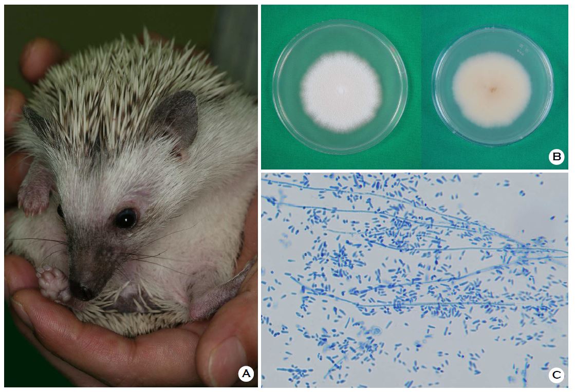
The pet hedgehog species, Atelerix albiventris, is characterized by four toes on its rear legs (Figure 1A). Macroscopically, T. erinacei has a white cottony appearance on the surface with a yellowish-brown pigmented undersurface (Figure 1B). Unlike typical T. mentagrophytes characteristics such as spiral hyphae and grape-shaped microconidia, T. erinacei has characteristic microconidia that are elongated, pear-shaped, and attached along the side of the hyphae (Figure 1C).
Macroconidia, when present, were slightly irregular in shape and size and have 2~6 septa. Reportedly, the combination of a white cottony surface and a yellow undersurface on Sabouraud's dextrose agar and pear-shaped microconidia is usually a distinctive characteristic of this species2,3. Microscopic examination and macroscopic morphology are crucial for identifying the causative organism; however, physicians can identify species more precisely using molecular biological analyses.
References
1. Perrier P, Monod M. Tinea manuum caused by Tricho- phyton erinacei: first report in Switzerland. Int J Dermatol 2015;54:959-960
Crossref
Google Scholar
PubMed
2. Rhee DY, Kim MS, Chang SE, Lee MW, Choi JH, Moon KC, et al. A case of tinea manuum caused by Trichophyton mentagrophytes var. erinacei: the first isolation in Korea. Mycoses 2009;52:287-290
Crossref
Google Scholar
3. Abarca ML, Castella G, Martorell J, Cabanes FJ. Trichophyton erinacei in pet hedgehogs in Spain: occurrence and revision of its taxonomic status. Med Mycol 2017;55:164-172
Crossref
Google Scholar
PubMed
Congratulatory MessageClick here!
