pISSN : 3058-423X eISSN: 3058-4302
Open Access, Peer-reviewed
pISSN : 3058-423X eISSN: 3058-4302
Open Access, Peer-reviewed

Eun Hye Jeong,Jeong Eun Yim,Hyeong Mok Kwon,Jong Soo Choi
10.17966/JMI.2022.27.1.23 Epub 2022 April 02
Abstract
PATIENT CONSENT STATEMENT: The patient provided written informed consent for the publication and the use of his images.
Keywords
Corkscrew pattern Dermoscopy Tinea capitis
Dermoscopy is a non-invasive diagnostic tool that visualizes the hair shafts, follicular ostia, and perifollicular skin to diagnose scalp and hair disorders. Although mycological examination is the gold standard for tinea capitis diagnosis, abnormal patterns of dermoscopic findings can help narrow the differential diagnosis1.
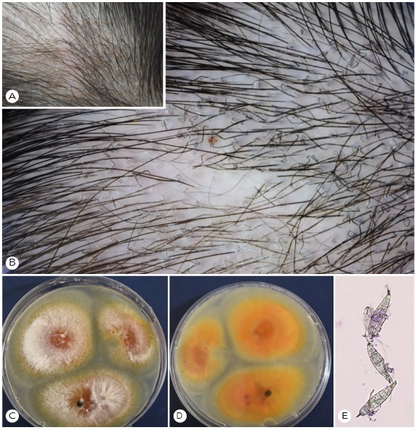
A one-year-old boy approached our clinic with a single scaly erythematous alopecic patch on his scalp. He had a history of contact with cats in the countryside, and the lesion occurred 15 days ago. On the background of the erythema, there were prominent corkscrew hair that were twisted and coiled observed during dermoscopy (Fig. 1A, 1B). On KOH examination, septated hyphae were found in the scalp scale, and spores were found on the outside of the hair shaft. On the Sabouraud agar plate, yellowish colonies grew quickly (Fig. 1C, 1D). The macroconidia had thick walls, more than six cells, and a curved knob-like end. These findings indicated the presence of Microsporum canis infection (Fig. 1E).
Comma, corkscrew, Morse code-like, zigzag, and bent hair are the most common dermoscopic findings of tinea capitis. Corkscrew hair were most noticeable in our case. Hughes et al. first described corkscrew hair in patients of African descent as a variation of comma-shaped hair2. Trichophyton and Microsporum species were found in cases of tinea capitis. According to the systemic review by Wakiel-Burnat et al., corkscrew hair has a sensitivity of 32% and a specificity of 100%3. It suggested that corkscrew hair is the specific indicator for diagnosing tinea capitis.
In conclusion, dermatologists need to consider that abnormal findings in dermoscopy can play an important role in the diagnosing tinea capitis. Herein, we report the corkscrew hair pattern upon dermoscopy, which was one of the specific findings.
References
1. Rudnicka L, Rakowska A, Kerzeja M, Olszewska M. Hair shafts in trichoscopy: clues for diagnosis of hair and scalp diseases. Dermatol Clin 2013;31:695-708
Google Scholar
2. Hughes R, Chiaverini C, Bahadoran P, Lacour JP. Cork- screw hair: a new dermoscopic sign for diagnosis of tinea capitis in black children. Arch Dermatol 2011;147: 355-356
Google Scholar
3. Waśkiel-Burnat A, Rakowska A, Sikora M, Ciechanowicz P, Olszewska M, Rudnicka L. Trichoscopy of tinea capitis: a systematic review. Dermatol Ther (Heidelb) 2020;10: 43-52
Google Scholar
Congratulatory MessageClick here!
