pISSN : 3058-423X eISSN: 3058-4302
Open Access, Peer-reviewed
pISSN : 3058-423X eISSN: 3058-4302
Open Access, Peer-reviewed

Seung-Min Oh,Hye-Jin Ahn,Min Kyung Shin
10.17966/JMI.2021.26.4.100 Epub 2022 January 01
Abstract
The patient provided written informed consent for the publication and the use of her images.
Keywords
Green nail syndrome Reflectance confocal microscopy
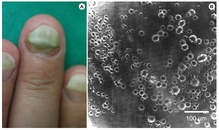
A 46-year-old female presented with greenish discoloration on fingernails. The patient had been diagnosed with onychomycosis at a local dermatology clinic. Physical examination revealed onycholysis on all fingernails and dark-greenish dis- coloration on the Lt 1st, 3rd and Rt 1st fingernail (Fig. 1A). KOH exam and reflectance confocal microscopy (RCM) images of the nail plate were all negative for fungal spores or hyphae. Multiple round, ring-shaped discs with white periphery and diameters ranging from 5 μm to 35 μm were observed in the RCM images (Fig. 1B). The patient was treated with oral ciprofloxacin and topical gentamycin and reported clinical improvement. Based on clinical and response of treatment, the diagnosis of green nail syndrome (GNS) was made. The causative agent is not definite without bacterial culture result, however, Pseudomonas aeruginosa is a reasonable candidate because it is the most common cause of GNS and susceptible to fluoroquinolones.
GNS is an infectious disorder of nails which presents green discoloration of the nail plate, proximal paronychia and distolateral onycholysis. Treatment options for GNS include oral fluoroquinolones, topical silver sulfadiazine, ciprofloxacin, and gentamicin1. RCM is a noninvasive method to evaluate fungal infection of nails by identifying bright, filamentous branching hyphae or roundish microspores2.
The diagnosis of GNS is mostly clinical but identification of bacterial colony on imaging tools such as RCM can aid in the diagnosis. Bjarnsholt et al. observed that in vivo biofilm patches mostly ranged from 5 to 200 μm in diameter3. The size of the discs in the patient's RCM image supports the notion that the discs represent bacterial biofilm formation on the patient's nail. Therefore, RCM images can provide with objective data that strengthen the diagnosis of GNS.
References
1. Chiriac A, Brzezinski P, Foia L, Marincu I. Chloronychia: green nail syndrome caused by Pseudomonas aeruginosa in elderly persons. Clin Interv Aging 2015;10:265-267
Google Scholar
2. Pharaon M, Gari-Toussaint M, Khemis A, Zorzi K, Petit L, Martel P, et al. Diagnosis and treatment monitoring of toenail onychomycosis by reflectance confocal micro- scopy: prospective cohort study in 58 patients. J Am Acad Dermatol 2014;71:56-61
Google Scholar
3. Bjarnsholt T, Alhede M, Alhede M, Eickhardt-Sørensen SR, Moser C, Kühl M, et al. The in vivo biofilm. Trends Microbiol 2013;21:466-474
Google Scholar
Congratulatory MessageClick here!
