pISSN : 3058-423X eISSN: 3058-4302
Open Access, Peer-reviewed
pISSN : 3058-423X eISSN: 3058-4302
Open Access, Peer-reviewed

Kyung Duck Park ,Weon Ju Lee
10.17966/JMI.2021.26.3.51 Epub 2021 October 01
Abstract
Dermatophytosis is a skin disorder caused by dermatophytes. Dermatophytes isolated in South Korea include Trichophyton (T.) rubrum, T. mentagrophytes, T. verrucosum, T. tonsurans, T. violaceum, T. schoenleinii, Microsporum (M.) canis, M. ferrugineum, M. gypseum, and Epidermophyton floccosum. T. tonsurans was first found in South Korea in 1992. In contrast, there have been no recent reported cases of T. violaceum, T. schoenleinii, and M. ferrugineum in South Korea. Population mobility, changes in human lifestyles, development of the healthcare system, and the introduction of antifungals have brought about dermatophyte evolution in the skin microenvironment. We have reviewed the cases of dermatophytosis caused by M. ferrugineum, T. violaceum, and T. schoenleinii reported both in South Korea and globally.
Keywords
Dermatophytosis Epidemiology Microsporum ferrugineum Trichophyton schoenleinii Trichophyton violaceum
Dermatophytosis is a skin disorder caused by dermatophytes. The genera of dermatophytic fungi include Trichophyton (T.), Microsporum (M.), and Epidermophyton (E.). The distribution of dermatophytes depends on geographical characteristics, economic conditions, and healthcare systems. Dermatophytes isolated in South Korea are T. rubrum, T. mentagrophytes, T. verrucosum, T. tonsurans, T. violaceum, T. schoenleinii, M. canis, M. ferrugineum, M. gypseum, and E. floccosum. T. tonsurans was first found in South Korea in 1992. Conversely, there have been no cases of T. violaceum, T. schoenleinii, and M. ferrugineum reported recently in South Korea. Bang and Lee reported data on dermatophytosis caused by M. ferrugineum in South Korea from 1976 to 1999. We have reviewed the epidemiological characteristics of M. ferrugineum infection in South Korea from 1976 to 2020. Furthermore, we have carried out a literature review to obtain data on dermatophytosis caused by T. violaceum and T. schoenleinii.
M. FERRUGINEUM INFECTION IS NO LONGER REPORTED IN SOUTH KOREA
M. ferrugineum is an anthropophilic dermatophyte that is mainly found in Africa, East Asia, and Eastern Europe. It often causes tinea capitis in children. M. ferrugineum fostered in culture media shows yellowish glabrous to downy colony growth. Lactophenol cotton-blue stain shows hyphae with prominent bamboo septa.
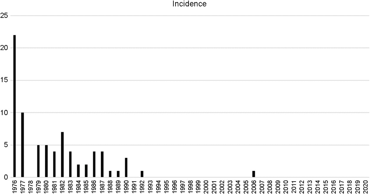
We gathered data from the medical records of 76 cases (55 men and 21 women) with M. ferrugineum infection collected by Kyungpook National University Hospital and the Catholic Skin Disease Clinic between 1976 and 2020. The incidence of M. ferrugineum infection declined in South Korea between 1976 and 2006 (Fig. 1). Since then, no more infections caused by M. ferrugineum were reported until 2020 (Fig. 1). In all, 97.4% of M. ferrugineum infection developed from 1976 to 1990 (Table 1). During this period, there was a considerably greater incidence of M. ferrugineum infection in children less than 10 years old (54.1%) than in any other age group. Tinea capitis was the most common clinical type (86.5%). There was only one reported case of M. ferrugineum infection between 1991 and 2005 (Table 1). The patient was one year old and had tinea faciei. Moreover, there was only one reported case of M. ferrugineum infection between 2006 and 2020 (Table 1). The patient was 27 years old and had tinea corporis. The last reported case of M. ferrugineum infection in South Korea was in 2006.
|
Year subtype Age |
1976~1990 |
|
1991~2005 |
|
2006~2020 |
Total |
|||
|
Tinea |
Tinea |
Tinea |
Tinea |
Tinea |
Tinea |
||||
|
0~9 |
37 |
1 |
2 |
|
|
1 |
|
|
41 |
|
10~19 |
24 |
2 |
2 |
|
|
|
|
|
28 |
|
20~29 |
|
|
|
|
|
|
|
1 |
1 |
|
30~39 |
1 |
|
1 |
|
|
|
|
|
2 |
|
40~49 |
|
|
|
|
|
|
|
|
0 |
|
50~59 |
1 |
|
|
|
|
|
|
|
1 |
|
60~69 |
1 |
|
|
|
|
|
|
|
1 |
|
70~79 |
|
|
|
2 |
|
|
|
|
2 |
|
Total |
64 |
3 |
5 |
2 |
|
1 |
|
1 |
76 |
|
Year Subtype |
1976~1990 |
1991~2005 |
2006~2020 |
Total |
|
|
|
|
|
|
|
Tinea capitis |
|
1 |
1 |
2 |
|
Tinea faciei |
1 |
2 |
1 |
4 |
|
Tinea corporis |
|
|
1 |
1 |
|
Tinea pedis |
|
|
|
|
|
Tinea unguium |
|
2 |
3 |
5 |
|
Total |
1 |
5 |
6 |
12 |
In addition, the Korean Journal of Dermatology reported four cases of M. ferrugineum infection. The patients were a nine-year-old boy with tinea corporis (reported in 1986), a two-year-old boy with tinea capitis (1992), a five-year-old boy with tinea capitis (1992), and a 10-year-old girl with tinea capitis (1992). The Journal of Mycology and Infection reported 27 cases of M. ferrugineum infection. These were seven patients with tinea pedis, four patients with tinea cruris, three patients with tinea manus, one patient with tinea faciei, and 12 patients with tinea capitis between 1976 and 1992. The other report of the Journal of Mycology and Infection showed that the proportion of M. ferrugineum infection in dermatophytosis was 0.6% in 1979, 0.5% in 1981, 6.2% in 1983, 0.2% in 1985, 0.4% in 1987, 0.2% in 1989, and 0.3% in 1992. According to this report, the last two patients (0.2%) with M. ferrugineum infection were in 1994. PubMed reported no cases of M. ferrugineum infection in South Korea.
M. FERRUGINEUM INFECTION IS STILL REPORTED GLOBALLY
In 2020, the German dermatologists Nenoff et al. reported that three boys developed tinea capitis caused by M. ferru- gineum. Moreover, M. ferrugineum was isolated in Germany in 2016. Nenoff et al. assumed that M. ferrugineum infection was a consequence of migration. Liang et al. described adult tinea capitis in China in 2019. They identified one patient with M. ferrugineum infection. In 2007, Ngwogu and Otokunefor reported the epidemiology of dermatophytosis in a rural community in Eastern Nigeria. In their study, M. ferrugineum was found in 7.3% of 4,287 primary school children with dermatophytosis.
DERMATOPHYTOSIS CAUSED BY T. VIOLACEUM IS NO LONGER REPORTED IN SOUTH KOREA
T. violaceum is an anthropophilic dermatophyte that is found mainly in Africa and the Middle East, in addition to parts of Europe. There are some endemic foci in South America and Mexico. T. violaceum is isolated primarily from tinea capitis, although it can infect glabrous skin, nails, and feet. T. violaceum grown on culture media shows a deep-red to violet glabrous colony. Lactophenol cotton-blue stain shows irregular hyphae without microconidia and macro- conidia.
We searched the Korean Journal of Dermatology, the Journal of Mycology and Infection, and PubMed for data on patients with T. violaceum infection. The Korean Journal of Dermatology reported a case of T. violaceum infection in South Korea in 1989. The patient was an eight-year-old with tinea faciei on the left lower eyelid. The Journal of Mycology and Infection reported a case of T. violaceum infection in 1996. The patient was an 82-year-old with tinea capitis and tinea faciei. The journal reported four other cases of T. violaceum infection in 2002: one patient had tinea capitis, one had tinea faciei, and two had onychomycosis. The journal reported another six cases in 2013; one case each of tinea capitis, tinea faciei, and tinea corporis, and three cases of tinea unguium (Table 2).
T. VIOLACEUM INFECTION IS ALSO EMERGING GLOBALLY
Gaviria Morales et al. conduced a retrospective analysis of dermatophytosis caused by T. violaceum in southern Switzerland from 2007 to 2018. Dermatophytosis due to T. violaceum was diagnosed in 44 patients in Switzerland. The authors assumed that people from endemic areas, mainly from Eritrea, were the main source of contagion. Zoulati et al. conducted a retrospective study of 12 cases of T. viola- ceum dermatophytosis in France between January 2011 and December 2016. Wiegand et al. examined the clinical presentation of tinea capitis in children in western Uganda. They found T. violaceum to be a causative agent for tinea capitis in 56.6% of the patients. Grigoryan et al. retro- spectively reviewed the charts of patients from Mayo Clinic in Rochester, Minnesota, United States, who had cultures positive for T. violaceum between 1997 and 2014. They concluded that T. violaceum is a tinea capitis pathogen that is most common among patients of African descent. Juncosa et al. reported all superficial mycosis cases caused by T. violaceum in patients receiving in-hospital treatment between 2000 and 2006. T. violaceum accounted for 18.5% of the 275 dermatophytes isolated during this period. Farina et al. evaluated the experience of an Italian multicenter with T. violaceum pathogens over a nine-year period (2005~2013). Twenty three strains were revealed as T. violaceum.
DERMATOPHYTOSIS CAUSED BY T. SCHOENLEINII IS NO LONGER REPORTED IN SOUTH KOREA
T. schoenleinii is an anthropophilic dermatophyte that is isolated mainly from certain regions of Eurasia and Africa. Though it has previously been found in certain small endemic areas in America, it has likely now been extirpated from either most or all of them. It is an agent of favus of the scalp and is characterized by the presence of scutula. T. schoenleinii grown on culture media shows a creamy glabrous to waxy colony. Lactophenol cotton-blue stain shows hyphae in the form of favic chandeliers without microconidia and macroconidia.
We searched the Korean Journal of Dermatology, the Journal of Mycology and Infection, and PubMed for data on patients with T. schoenleinii infection. The Journal of Mycology and Infection reported two cases of T. schoenleinii infection in 1979. The patients were a 17-year-old man and a 15-year-old woman with favus. The Korean Journal of Dermatology reported three cases of T. schoenleinii infection in 1987 (Table 3).
|
Year Subtype |
1976~1990 |
1991~2005 |
2006~2020 |
Total |
|
Tinea capitis |
4 |
|
|
4 |
|
Tinea faciei |
|
|
|
|
|
Tinea corporis |
|
|
|
|
|
Tinea pedis |
1 |
|
|
1 |
|
Tinea unguium |
|
|
|
|
|
Total |
5 |
|
|
5 |
T. SCHOENLEINII INFECTION IS RARELY REPORTED GLOBALLY
There have been very few reports of favus over the past four decades. Interestingly, Iwasa et al. reported a case of favus of vellus hair due to T. schoenleinii in a 63-year-old Japanese woman in 2019. Mansouri et al. reported a case of extensive tinea corporis in an 80-year-old woman in Iran in 2012. Ghadgepatil et al. identified an unusual case of tinea capitis due to T. schoenleinii in an elderly woman in India in 2015. Other cases have been reported from Tunis in 2007, from Romania in 2012, and from Poland in 2012.
M. ferrugineum, T. violaceum, and T. schoenleinii were once major pathogens of dermatophytosis. However, since the mid-twentieth century their incidence has decreased dramatically, and they are now endemic in some less-developed countries. Conversely, the incidences of T. rubrum, T. interdigitale, T. tonsurans, and M. canis have increased gradually, and these fungi have become the major species worldwide. Currently, T. rubrum is the leading pathogen of dermatophytosis, and M. canis and T. tonsurans are the predominant dermatophytes of tinea capitis. The incidence of dermatophytosis in South Korea has shown the same trend as that of the global incidence. According to Lee et al., the annual incidence of patients with T. rubrum infection in South Korea has increased over the past 37 years and 88.35% of patients who presented with dermatophytosis had T. rubrum infection. T. mentagrophytes is the second most common pathogen of dermatophytosis in South Korea, and M. canis is the third. Population mobility, changes in human lifestyles, development of the healthcare system, and emergence of antifungal agents will continually drive the evolution of dermatophytes in the skin microenvironment. M. ferrugineum, which was once a representative strain of tinea capitis in- fection, has not been reported from South Korea since 2006. This is thought to be due to rapid changes in socioeconomic status, including economic development and improved living standards. T. schoenleinii is known to cause favus, and there have been many reports since the first report in South Korea in 1954; however, there have been no reports since 1987. This trend is thought to be related to the social chaos resulting from the Korean War from 1950 to 1953 and the rapid population change. According to Suh, T. schoenleinii was introduced by refugees from either North Korea or China during the Korean War. Relatively than pre- vious two species, T. violaceum has been reported in South Korea recently, and has been reported again in Europe. Thus, this species needs to be continuously confirmed. The limitations of this study include the possibility that we missed cases. Moreover, we did not cover data reported before the 1970s. In conclusion, there is a need for comprehensive observation to understand such fungi better and anticipate future trends and changes.
References
1. Bang YJ, Lee GS. The epidemic study on Microsporum ferrugineum. Korean J Clin Lab Sci 2000;32:62-65
Google Scholar
2. Lee KH, Lee ES, Kang WH, Lee SN. An unusual clinical manifestation of tinea corporis caused by Microsporum ferrugineum. Korean J Dermatol 1987;25:383-389
Google Scholar
3. Kim HU, Choi CJ, Yun SK. Three cases of tinea capitis caused by Microsporum ferrugineum. Korean J Dermatol 1993;31:760-765
Google Scholar
4. Ahn KJ, Jang SJ. Superficial dermatomycosis and the causative agents in Korea. KJMM 2004;9:91-99
Google Scholar
5. Han ES, Seo SJ, Kim MN, Hong CK, Ro BI. A clinical and mycological study of superficial fungal diseases (VIII). Korean J Med Mycol 1996;1:91-100
Google Scholar
6. Nenoff P, Gebhardt M, Klonowski E, Koch D, Krüger C, Uhrlaß S. Microsporum ferrugineum-an anthropophilic dermatophyte in Germany: case report and review of the literature. Hautarzt 2020;71:705-710
Google Scholar
7. Liang G, Zheng X, Song G, Zhang M, Liu J, Zang X, et al. Adult tinea capitis in China: a retrospective analysis from 2000 to 2019. Mycoses 2020;63:876-888
Google Scholar
8. Ngwogu AC, Otokunefor TV. Epidemiology of dermato- phytoses in a rural community in Eastern Nigeria and review of literature from Africa. Mycopathologia 2007; 164:149-158
Google Scholar
9. Kim YA, Lee KH, Lee JB, Suh SB. A case of fungal granu- loma caused by Trichophyton violaceum. Korean J Dermatol 1989;27:304-308
Google Scholar
10. Lee JB, Kwon KS, Chung TA, Jang HS, Oh CK. A case of black dot ringworm caused by Trichophyton violaceum. Korean J Med Mycol 1998;3:39-42
Google Scholar
11. Moon HJ, Lee JB, Kim SJ, Lee SC, Won YH. Clinical and mycological studies on dermatomycosis (1991-2000). Korean J Med Mycol 2002;7:78-85
Google Scholar
12. Oh SJ, Lee SY, Lee JS. A clinical and mycological study of dermatophytoses in Chungcheongnam-do Province (2008~2012). KJMM 2013;18:39-47
Google Scholar
13. Gaviria Morales E, Iorizzo M, Martinetti Lucchini G, Mainetti C. Trichophyton violaceum: an emerging patho- gen in Southern Switzerland. Dermatology 2019;235: 434-439
Google Scholar
14. Zoulati G, Maïga RY, El Haouri M, Er-Rami M. Der- matophytosis due to Trichophyton violaceum at the parasitology-mycology laboratory of the military hospital of Meknes (about twelve cases). J Mycol Med 2018;28: 1-7
Google Scholar
15. Wiegand C, Mugisha P, Mulyowa GK, Elsner P, Hipler UC, Gräser Y, et al. Trichophyton violaceum: main cause of tinea capitis in children at Mbarara Regional Referral Hospital in Uganda. Hautarzt 2016;67:712-717
16. Grigoryan KV, Tollefson MM, Olson MA, Newman CC. Pediatric tinea capitis caused by Trichophyton violaceum and Trichophyton soudanense in Rochester, Minnesota, United States. Int J Dermatol 2019;58:912-915
Google Scholar
17. Juncosa T, Aguilera P, Jaen A, Vicente A, Aguilar AC, Fumadó V. Trichophyton violaceum: an emerging patho- gen. Enferm Infecc Microbiol Clin 2008;26:502-504
Google Scholar
18. Farina C, Fazii P, Imberti G, Lombardi G, Passera M, Andreoni S, et al. Trichophyton violaceum and T. sou- danense: re-emerging pathogens in Italy, 2005-2013. New Microbiol 2015;38:409-415
Google Scholar
19. Kim KH, Bang YJ, Jun JB, Kim H. Favus diagnosed in siblings in 1979. Korean J Med Mycol 2017;22:178-181
Google Scholar
20. Won YH, Kim SH, Kim SH, Kim YP. A clinical and mycological studies of dermatomycosis (1976~1985). Korean J Dermatol 1987;25:753-761
Google Scholar
21. Iwasa K, Ogawa K, Azukizawa H, Tanabe H, Iwanaga T, Anzawa K, et al. Revival of favus in Japan caused by Trichophyton schoenleinii. J Dermatol 2019;46:347-350
Google Scholar
22. Mansouri P, Farshi S, Khosravi AR, Naraghi ZS, Chalangari R. Trichophyton schoenleinii-induced widespread tinea corporis mimicking parapsoriasis. J Mycol Med 2012;22: 201-205
Google Scholar
23. Ghadgepatil SS, Sharma YK, Misra R, Dash KN, Patvekar MA, Deo KS. An unusual case of tinea capitis caused by Trichophyton schoenleinii in an elderly female. Indian Dermatol Online J 2015;6:49-50
Google Scholar
24. Khaled A, Ben Mbarek L, Kharfi M, Zeglaoui F, Bouratbine A, Fazaa B, et al. Tinea capitis favosa due to Trichophyton schoenleinii. Acta Dermatovenerol Alp Pannonica Adriat 2007;16:34-36
Google Scholar
25. Mareş M, Năstasă V, Apetrei IC, Suditu GC. Tinea corporis bullosa due to Trichophyton schoenleinii: case report. Mycopathologia 2012;174:319-322
Google Scholar
26. Macura AB, Krzyściak P, Skóra M, Gniadek A. Case report: onychomycosis due to Trichophyton schoenleinii. Mycoses 2012;55:e18-19
Google Scholar
27. Zhan P, Liu W. The changing face of dermatophytic in- fections worldwide. Mycopathologia 2017;182:77-86
Google Scholar
28. Lee WJ, Kim SL, Jang YH, Lee SJ, Kim DW, Bang YJ, et al. Increasing prevalence of Trichophyton rubrum identified through an analysis of 115,846 cases over the last 37 years. J Korean Med Sci 2015;30:639-643
Google Scholar
29. Kim SL, Lee KC, Jang YH, Lee SJ, Kim DW, Lee WJ, et al. The epidemiology of dermatophyte infection in South- eastern Korea (1979~2013). Ann Dermatol 2016;28: 524-527
Google Scholar
30. Suh SB. Dermatophytosis and its causative agents in Korea. Korean J Med Mycol 1996;1:1-10
Google Scholar
31. Kim KH. Changing patterns of dermatophytosis and its causative agents according to social and economic devel- opments in Korea. Korean J Med Mycol 2006;11:1-12
Google Scholar
Congratulatory MessageClick here!
