pISSN : 3058-423X eISSN: 3058-4302
Open Access, Peer-reviewed
pISSN : 3058-423X eISSN: 3058-4302
Open Access, Peer-reviewed

Jeongsoo Lee,Nuri Na,Joonsoo Park
10.17966/JMI.2020.25.1.26 Epub 2020 April 07
Abstract
Keywords
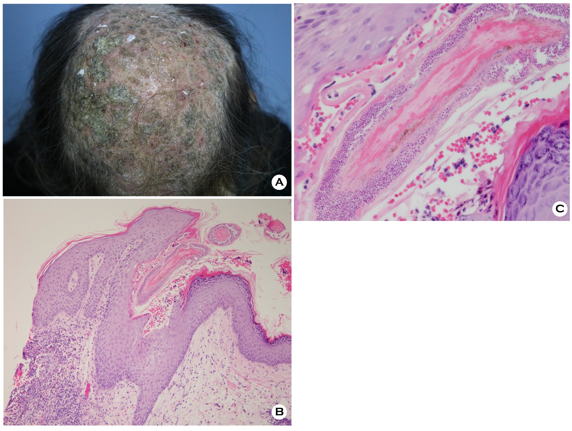
Tinea capitis is a scalp infection caused by dermatophytes, primarily Trichophyton and Microsporum spp.1 Kerion celsi is a severe, boggy inflammatory form of tinea capitis. Deep, tender, and boggy plaques with pus are known clinical features of kerion celsi2. The patient may develop single or multiple round-shaped patches of hair loss with scarring in the areas of inflammation and suppuration (Figure 1A).
Kerion celsi is commonly confused with bacterial abscess because of purulent drainage. It must be clinically differentiated from chronic staphylococcal folliculitis, pediculosis capitis, psoriasis, seborrheic dermatitis, and various inflammatory follicular conditions2.
Histologically well-established lesions of kerion celsi show foci of parakeratosis with epidermal acanthosis and spongiosis. In the dermal and perifollicular area, severe inflammatory tissue reactions can be observed, such as neutrophil infiltration (Figure 1B). Numerous fungal spores surrounding the hair shaft and follicle represent a distinct specific histological feature in kerion celsi (Figure 1C).
Fungal culture is the gold standard method to confirm the fungal infection. However, patients with kerion celsi show a high rate of false-negative results with conventional culture methods, chiefly because the samples contain inflammatory cells without fungal cells1. Histological analysis is a successful alternative method in culture-negative patients for identifying fungus without special stains, such as periodic acid-Schiff and Grocott's methenamine silver.
The patient provided written informed consent for the publication and the use of his or her images.
References
1. John AM, Schwartz RA, Janniger CK. The kerion: an angry tinea capitis. Int J Dermatol. 2018;57:3-9
Google Scholar
2. James WD, Berger TG, Elston DM. Andrews' diseases of the skin. 12th ed. Philadelphia: Elsevier, 2015:286-294
Congratulatory MessageClick here!
