pISSN : 3058-423X eISSN: 3058-4302
Open Access, Peer-reviewed
pISSN : 3058-423X eISSN: 3058-4302
Open Access, Peer-reviewed
Yong Woo Choi,Joonsoo Park,Hyun Hee Kwon
http://dx.doi.org/10.17966/KJMM.2017.22.4.188 Epub 2017 December 22
Abstract
Keywords
Aspergilloma Aspergillosis Fungus ball
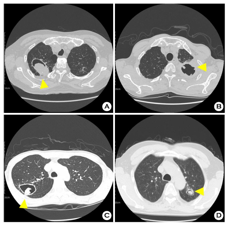
Aspergillosis is a mycotic disease caused by Aspergillus species, usually Aspergillus fumigatus[1]. Saprophytic aspergillosis, so called aspergilloma, is characterized by Aspergillus infection without other tissue invasion[2]. Aspergilloma typically leads to conglomeration of interrelated fungal hyphae admixed with mucus and cellular debris within a preexistent pulmonary cavity or bronchus[2]. At computed tomography (CT), mycetomas are characterized by the existence of a solid, round or oval mass with soft-tissue opacity within a lung cavity[2].
The "air crescent" sign is the mass that separated from the wall of the cavity by an airspace of variable size and shape[3]. Other causes of the air crescent sign include angioinvasive aspergillosis, echinococcal cyst, and rarely, tuberculosis, tuberculosis cavity, lung abscess, bronchogenic carcinoma, and P. carinii pneumonia[3]. The mass is typically spherical or ovoid, and usually moves when the patient changes position[2],[3]. In axial sections of CT scan shows a relatively thin walled cavity containing a hyperdense soft tissue accentuation with surrounding air lucency. Few fibrotic changes are seen surrounding the cavity (Fig. 1-A, B, C, D).
Aspergillosis is a serious complication that is frequently seen in immunocompromised patients[2]. Although CT findings in various type of pulmonary aspergillosis may be nonspecific, in the appropriate clinical setting, familiarity with the thin-section CT findings may help establish the specific diagnosis.
References
1. Franquet T, Müller NL, Giménez A, Guembe P, de La Torre J, Bagué S. Spectrum of pulmonary Aspergillosis: Histologic, cinical, and radiologic findings. Radiographics 2001;21:825-837
Crossref
Google Scholar
2. Godet C, Laurent F, Bergeron A, Ingrand P, Beigelman-Aubry C, Camara B, et al. CT imaging assessment of response to treatment in chronic pulmonary aspergillosis. Chest 2016;150:139-147
Crossref
3. Raju S, Ghosh S, Mehta AC. Chest CT sighs in pulmonary disease: A pictorial review. Chest 2017; 151:1356-1374
Google Scholar
