pISSN : 3058-423X eISSN: 3058-4302
Open Access, Peer-reviewed
pISSN : 3058-423X eISSN: 3058-4302
Open Access, Peer-reviewed
Yong Woo Choi,Osung Kwon,Hyun Chung ,Joonsoo Park
http://dx.doi.org/10.17966/KJMM.2017.22.3.146 Epub 2017 September 29
Abstract
Keywords
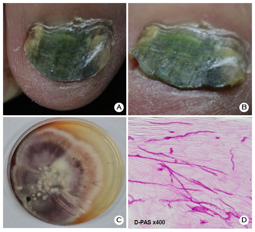
Chromonychia Green nail syndrome Pseudomonas aeruginosa
Green nail syndrome (GNS) is characterized by greenish chromonychia caused by pyocyanin, a metabolite produced by Pseudomonas aeruginosa (P. aeruginosa)[1]. Predisposing factors of pseudomonal colonization include onychomycoses, nail dystrophies, working in wet conditions, diabetes mellitus, paronychia and immunosuppressive states[2]. A number of reports states that there are strong relationship between fungal and P. aeruginosa infection of the nail[3],[4]. Fungal infection stimulates bacterial colonization within the nail and overgrowth of P. aeruginosa[4].
References
1. Agger WA, Mardan A. Pseudomonas aeruginosa infections of intact skin. Clin Infect Dis 1995;20:302 -308
Crossref
Google Scholar
2. Maes M, Richert B, de la Brassinne M. Green nail syndrome or chloronychia. Rev Med Liege 2002;57: 233-235
Google Scholar
3. Moore M. Green nails; the role of Candida (syrin- gospora, Monilia) and Pseudomonas aeruginosa. AMA Arch Derm Syphilol 1951;64:499-505
Google Scholar
4. Pier GB, Ramphal R. Pseudomonas aeruginosa. Principles and practive of infectious diseases. 6th ed. Philadelphia, PA, USA: Churchill Livingstone; 2004: 2834-2860
