pISSN : 3058-423X eISSN: 3058-4302
Open Access, Peer-reviewed
pISSN : 3058-423X eISSN: 3058-4302
Open Access, Peer-reviewed

YongWoo Choi,Nuri Na,Jong Soo Choi,Joonsoo Park
10.17966/JMI.2019.24.2.66 Epub 2019 July 03
Abstract
Keywords
Conidiophore Pseudallescheria boydii Scedos- porium apiospermum
Pseudallescheria boydii (P. boydii) is a species of fungus classified in the Ascomycota1. Formerly known as Monosporium apiospermum, Scedosporium apiospermum is the anamorph of P. boydii1. Typically found in stagnant and polluted water, P. boydii has been implicated in the infection of immunocompromized and near-drowned pneumonia patients2. Diagnosis of P. boydii is possible through isolation of the fungus in culture or through cytology and histopathology in the tissues of diseased individuals3. The treatment of P. boydii infections is complicated by its resistance to many standard antifungal agents used to treat infections by filamentous fungi3.
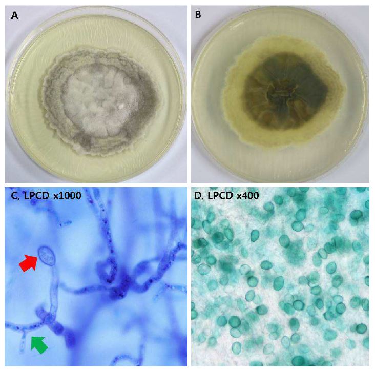
On Saboraud Dextrose Agar, P. boydii colonies mature rapidly and are flat, white colored, and texture ranges from wooly to cottony1. The colonies become grayish or smoky-brown colored with age (Figure 1A); some laboratorians refer to a "house mouse gray" color3. The color of the colony is white initially but darkens to gray to black with maturity on the reverse side (Figure 1B). Microscopically, the conidia of P. boydii are unicellular, pale brown, and ovoid with truncated bases formed singly, in small clusters at the ends of the conidiophores, or from short annelid necks arising directly from the hyphae3. Conidiophores bearing annelids are of varying length and exhibit little differentiation from the vegetative hyphae1. Fascicles of conidiophores bound together in synemata are sometimes present and are a variation of the asexual state referred to as the Graphium synanamorph3. Large (50~ 250 μm) brown cleistiothecia of the sexual P. boydii may develop after 2~3 weeks of incubation and are likely to be found at the center of a colony3. Ascospores are yellow-brown and ellipsoidal in shape3.
References
1. Karen C, Michael A, Marie L, Alexander J, Robin P, Sandra S, et al. Manual of clinical microbiology. 12th ed. Wachington, DC: ASM Press, 2019:2007-2305
2. Guarro J, Kantarcioglu AS, Horré R, Rodriguez-Tudela JL, Cuenca E, Berenguer J, et al. Scedosporium apiospermun: changing clinical spectrum of a therapy-refractory opportunist. Med Mycol 2006;44:295-327
Google Scholar
3. Karoll J, Emmanuel R, Flavio Q, Joseph M, Charalampos A, Tena K, et al. Infections caused by Scedosporium spp. Clin Microbiol Rev 2008;21:157-197
Google Scholar
Congratulatory MessageClick here!
