pISSN : 3058-423X eISSN: 3058-4302
Open Access, Peer-reviewed
pISSN : 3058-423X eISSN: 3058-4302
Open Access, Peer-reviewed

Hye Ri Kim,Dong Hoon Shin,Jee-Bum Lee,Jong Soo Choi
10.17966/JMI.2019.24.2.45 Epub 2019 July 03
Abstract
Background: Sporotrichosis is a common deep mycosis caused by the Sporothrix schenckii complex. Until 2016, no molecular studies had been conducted on these fungi, and all the included strains were reported as S. schenckii. However, investigations conducted in northeast China, Japan, and India revealed that S. globosa was the most prevalent Sporothrix species, whereas S. schenckii sensu stricto was reported very rarely.
Objective: To investigate the accurate prevalent causative species of sporotrichosis among strains reported as S. schenckii in Korea.
Methods: We isolated strains of Sporothrix spp. Prevalent in Korea from fungus collection centers or private collections and reviewed the available literature on molecular studies of strains from this region. We found five S. schenckii (1998-2016) and three S. globosa (2016-2018) strains. Ribosomal DNA internal transcribed spacer (ITS) sequences of these strains were compared with those of the S. schenckii complex strains.
Results: The ribosomal ITS sequences of the eight strains were 100% identical with that of S. globose. No S. schenckii sensu stricto was found. In addition, a study on the molecular analysis of Korean S. schenckii published by Ishizaki et al. (2004) demonstrated that the eight strains were of the mitochondrial subtype group B (S. globosa). Thus, all the 16 strains examined within the Korean S. schenckii complex were determined to be S. globosa.
Conclusion: In summary, S. globosa is the causative species within the tested Korean sporotrichosis cases reported between 1998 and 2018. Based on our analyses, S. globosa, and not S. schenckii, may be the predominant species in Korea.
Keywords
Sporotrichosis Sporothrix globosa
Sporotrichosis is a subcutaneous or systemic fungal infection caused by the Sporothrix schenckii complex. Under ordinary circumstances, Sporothrix infection occurs via traumatic inoculation of the skin or subcutis. S. schenckii has been considered the sole species causing all types of sporotrichosis in humans1. In 2007, Marimon et al.2 reported the following six species of S. schenckii complex based on phenotypic and genotypic analyses: S. schenckii sensu stricto, S. globosa, S. brasiliensis, S. luriei, S. mexicana, and S. pallida (S. albicans). Ishizaki et al.3 investigated 357 Japanese isolates of Sporothrix via restriction fragment length polymorphism (RFLP) analysis of mitochondrial DNA (mtDNA) and discovered only 15 isolates belonging to group A (S. schenckii; 4.2%), with all other isolates belonging to group B (S. globosa; 342/357, 95.8%). Liu et al.4 and Yu et al.5 demonstrated that in northeast China, all of the sporotrichosis-causing S. schenckii complex strains were S. globosa.
Since 1970, a total of 521 cases of sporotrichosis were published within 31 reports in Korea, and all those cases were reportedly caused by S. schenckii6-36. No molecular studies were conducted until 2016, and S. schenckii was long believed to be the causative species of sporotrichosis in Korea. Kim et al.37,38 reported three cases of human sporotrichosis caused by S. globosa and suggested that this fungus is the only causative organism of sporotrichosis in Korea. To clarify the causative pathogen, the current study aimed to collect strains of Sporothrix prevalent in Korea and analyze them using ribosomal DNA internal transcribed spacer (ITS) sequences.
1. Fungal isolates
This study included five and three clinical isolates reported as S. schenckii (1998-2016) and S. globosa (2016-2018), respectively, in Korea (Table 1). These strains were isolated from fungus collection centers or private collections. The isolates were cultured on potato dextrose agar (PDA; DifcoTM Becton, Dickinson and Company, Sparks, USA) and incubated at -80℃. In addition, eight Korean strains described before in 2003 by Ishizaki et al. were included39 (Table 2).
|
No |
Year |
Area |
Source |
Reported
as |
rDNA ITS |
|
1 |
1998 |
Gwangju |
Human
skin |
S. schenckii |
S. globosa |
|
2 |
2009 |
Gwangju |
Human
skin |
S. schenckii |
S. globosa |
|
3 |
2009 |
Gwangju |
Human
skin |
S. schenckii |
S. globosa |
|
4 |
2015 |
Gyeungju |
Human
skin |
S. schenckii |
S. globosa |
|
5 |
2016 |
Gyeungju |
Human
skin |
S. schenckii |
S. globosa |
|
6 |
2016 |
Daegu |
Human
skin |
S. globosa |
S. globosa |
|
7 |
2018 |
Daegu |
Human
skin |
S. globosa |
S. globosa |
|
8 |
2018 |
Daegu |
Human
skin |
S. globosa |
S. globosa |
|
No |
Year |
Area |
Source |
Reported as |
mtDNA type |
rDNA ITS |
|
1 |
<2003 |
Korea |
Human skin |
S. schenckii |
4/B |
S. globosa |
|
2 |
<2003 |
Korea |
Human skin |
S. schenckii |
4/B |
S. globosa |
|
3 |
<2003 |
Korea |
Human skin |
S. schenckii |
4/B |
S. globosa |
|
4 |
<2003 |
Korea |
Human skin |
S. schenckii |
4/B |
S. globosa |
|
5 |
<2003 |
Korea |
Human skin |
S. schenckii |
4/B |
S. globosa |
|
6 |
<2003 |
Korea |
Human skin |
S. schenckii |
5/B |
S. globosa |
|
7 |
<2003 |
Korea |
Human skin |
S. schenckii |
5/B |
S. globosa |
|
8 |
<2003 |
Korea |
Human skin |
S. schenckii |
5/B |
S. globosa |
2. DNA extraction and amplification
The Sporothrix spp. strains were sub-cultured on PDA for at least 2 weeks at 28℃. Then, genomic DNA was directly extracted and purified from the fungal colonies using the QIAmp DNA Mini Kit (Qiagen, Hilden, Germany). Approximately 1 mg of fresh filamentous mycelia were scraped and incubated overnight at 56℃ in 1.5 mL microcentrifuge tubes containing 180 μL of animal tissue lysis buffer and 20 μL of proteinase K. Additional lysis buffer (200 μL) was added and mixed by pulse vortexing for 15 s, and the solution was then incubated at 70℃ for 10 min. Next, 200 μL of ethanol was added to the sample after which it was vortexed and briefly centrifuged. The sample mix was then carefully added to a QIAmp Mini column and centrifuged for 1 min. After switching on the vacuum pump, wash buffer (AW1 and AW2; 500 μL each) was added and the sample was centrifuged twice. Subsequently, 100 μL of elution buffer was added to the sample mixt, after which the sample was incubated for 5 min at room temperature (20~25℃) and centrifuged for 1 min. The total volume of the final extraction product was approximately 100 μL, which was preserved at -20℃ until use.
DNA was amplified using polymerase chain reaction (PCR) with the primer pair ITS1 (5'-TCCGTAGGTGAACCTGCGG-3') and ITS4 (5'-TCCTCCGCTTATTGATATGC-3'). The PCR mixture consisted of 15 μL of 2× PCR premix, 1 μL of the DNA template, 2 μL of ultrapure water, and 1 μL of each primer (10 pmol/μL). The cycling conditions were as follows: 5 min at 95°C for initial denaturation; 30 cycles of 30 s at 95℃, 30 s at 60℃, and 1 min at 72℃ for amplification; and 10 min at 72℃ for extension. The PCR product quantities were determined using agarose gel electrophoresis, and the products were then purified using a purification kit (Bioneer, Daejoon, Korea).
3. Sequencing
Automated sequencing was performed at Macrogen (Seoul, Korea), and ribosomal DNA (rDNA) ITS sequences were compared with those of the the S. schenckii complex strains using BLAST®.
4. Phylogenetic study
The ITS sequences of the Korean isolates were compared with those of the S. schenckii complex strains available in GenBank. Evolutionary history was inferred using the neighbor- joining method in MEGA X40-43. Evolutionary distances were computed using the Kimura 2-parameter method. All positions containing gaps and missing data were eliminated (complete deletion option).
The ribosomal ITS sequence BLAST search revealed that all the five strains initially reported as S. schenckii and the three strains of S. globosa were 100% identical to S. globosa. S. schenckii sensu stricto was not detected. The phylogenetic tree of the rDNA ITS sequences showed that six well-defined pathogenic species and all clinical isolates from South Korea were clustered into the S. globosa group (Figure 1).
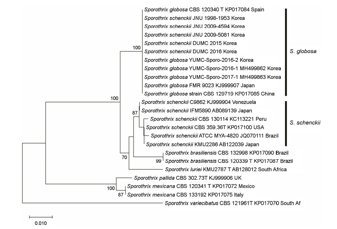
The mtDNA analysis of eight S. schenckii strains isolated from South Korea by Ishizaki et al.39 in 2004 were identified as being mitochondrial type 4 and 5, which belong to group B (S. globosa). Therefore, our molecular identification revealed that all 16 strains of the Korean S. schenckii complex actually belonged to S. globosa.
Sporotrichosis is a common deep mycosis and was previously considered to be caused solely by the S. schenckii complex strains. However, with the decline in the rural population and improvements in mechanized agriculture and personal hygiene due to industrialization, the incidence of sporotrichosis cases has been decreasing10.
In 2000, Sporothrix isolates were classified into 24 mtDNA types (types 1~24) based on RFLP patterns, and Ishizaki et al.3 classified these types phylogenetically into Groups A (types 1~3, 11, 14~19, 22, and 23) and B (types 4~10, 12, 13, 20, 21, and 24). Later, using molecular analyzes, such as ITS, and markers, such as β-tubulin, chitin synthase, and calmodulin, S. schenckii was identified as a species complex comprising the species S. schenckii sensu stricto, S. globosa, S. luriei, S. brasiliensis, S. mexicana, and S. pallida44. Analysis of the phenotypic characteristics and molecular identities revealed that S. schenckii sensu stricto, S. globosa, S. luriei, and S. brasiliensis, which are grouped into S. schenckii sensu lato, show phylogenetic proximity with each other, whereas S. mexicana and S. pallida are placed in highly remote positions. Furthermore, the phylogenetic tree indicated that S. mexicana and S. pallida share a common ancestor (Figure 1)44.
Zhang et al.45 reviewed the literature data from Asia-including China, India, and Japan-wherein there were 4,275 reported cases of sporotrichosis, whereas sequenced isolates comprised of only 140 strains. Of those, 139 (99.3%) were classified as S. globosa, and only one was identified as S. schenckii. In Japan, Ishizaki et al.3 analyzed 357 Sporothrix isolates using RFLP analysis of mtDNA and revealed that most species (342/357) belonged to group B (S. globosa) and very few (15/357) belonged to group A (S. schenckii). More than 4,000 cases of sporotrichosis were reported throughout most of the Chinese provinces, with the largest number of cases occurring in northeast China46. S. globosa (685/689, 99.4%) was the most prevalent, and S. schenckii (4/689, 0.6%) was very rare in China47. In India, sporotrichosis cases have been reported to cluster within the sub-Himalayan region in the northeast states, and the 14 sequenced isolates were all found to be S. globosa46,47. The number of reported cases and sequenced isolates are listed in Table 3. In Asia, S. globosa is the predominant endemic species, whereas S. schenkii is extremely rare and S. brasiliensis has not been reported47.
|
Nation |
Reported cases |
Sequenced isolates (%) |
||
|
Total |
S. globosa |
S. schenckii |
||
|
Japan |
555 |
42 |
41 (97.6) |
1 (2.4) |
|
3573 |
342 (95.8) |
15 (4.2) |
||
|
China |
4,482 |
689 |
685 (99.4) |
4 (0.6) |
|
India |
2,983 |
14 |
14 (100) |
0 (0) |
|
Korea |
521 |
16 |
16 (100) |
0 (0) |
In Korea, prior to 2016, based on morphological studies, S. schenckii was reportedly the causative species of sporotrichosis6-36. Recent case reports by Kim et al.37,38 in South Korea revealed three causative organisms to be S. globosa by molecular studies. To confirm the causative species of sporotrichosis within South Korea, we collected strains and reviewed the literature39. Our study, to some extent, identified sixteen causative species as S. globosa using rDNA ITS sequencing and mtDNA types.
S. globosa is endemic to Asia, and Moussa et al.47 suggested a hypothesis of an India-China-Japan belt for S. globosa prevalence. Our research supports the statistical probability that S. globosa is the predominant species in Korea and that Korea should also be included in this proposed belt45-47.
The limitations of the current study include the small sample size (16 strains), collection within a short period of time (1998-2018), and the lack of causative species sourced from the environment. Further studies examining more Sporothrix strains from fungus collection centers should be conducted. Because sporotrichosis commonly occurs in mammals mostly through "sapronosis" (caused by organisms of the environment rather than a living host) or "zoonosis" (transmitted from animals to humans), there is also a need to investigate and isolate Sporothrix spp. from surrounding environments48. Studies integrating other human pathogens as well as environmental species should be performed.
To conduct a comprehensive overview of the causative species of sporotrichosis, we need to sequence all the strains that have been reported in Korea. Despite 521 cases being reported in Korea, only eight strains were adequately stored, whereas most of them were discarded. To perform more optimal studies in the future, fungal strains should be collected and stored within well-controlled settings for adequate long-term preservation. Therefore, a collection center that guarantees a state of quality and maintenance of all regulations concerning biosafety is urgently needed in Korea.
In Brazil, S. brasiliensis reportedly caused an epidemic zoonosis affecting domestic cats44. New causative species, such as S. braziliensis, could be imported into Korea due to globalization. Therefore, a range of sporotrichosis infections, particularly those in cats, should be monitored.
In conclusion, as previously considered, S. schenckii is not a unique causative species of sporotrichosis in Korea. We identified that the 16 strains of sporotrichosis reported in Korea since 1998 belonged to S. globosa. Nowadays, there are several methods available to confirm sporotrichosis species, such as direct morphological examination, fungal culture, and molecular biology diagnosis44. Thus, it is critical that we carefully observe and examine samples from Sporothrix infections.
References
1. Rippon JW. Medical mycology: the pathogenic fungi and the pathogenic actinomycetes. 3rd ed. Philadelphia: WB Saunders, 1988:325-328
Google Scholar
2. Marimon R, Cano J, Gene J, Sutton DA, Kawasaki M, Guarro J. Sporothrix brasiliensis, S. globosa and S. mexicana, three new Sporothrix species of clinical interest. J Clin Microbiol 2007;45:3198-3206
Google Scholar
3. shizaki H, Kawasaki M. Molecular epidemiology of Sporothrix schenkii. Nihon Ishinkin Gakkai Zasshi 2000;41:245 -249
Google Scholar
4. Liu TT, Zhang K, Zhou X. Molecular identification of Sporothrix clinical isolates in China. J Zhejiang Univ Sci B 2014;15:100-108
Google Scholar
5. Yu X, Wan Z, Zhang Z, Li F, Li R, Liu X. Phenotypic and molecular identification of Sporothrix isolates of clinical origin in northeast China. Mycopathologia 2013;176:67-74
Google Scholar
6. Yoon S, Sihn JY, Kim YP. Clinical analysis of twelve cases of sporotrichosis: report of two cases and ten cases appeared in literatures of Korea. Korean J Dermatol 1972; 10:171-177
Google Scholar
7. Ihm CW. Mycotic diseases among farmers and fisheries in Korea: II. clinical, mycological, histopathological and epidemiological studies of twenty six cases of cutaneous sporotrichosis. Korean J Dermatol 1972;10:137-152
Google Scholar
8. Kim KH, Jeon US, Suh SB. A case of sporotrichosis developed after double eyelid operation. Korean J Dermatol 1975;13:193-198
Google Scholar
9. Hyun GM, Park YC, Kim JT. A case of sporotrichosis. Korean J Dermatol 1976;14:147-152
10. Kim BS, Suh SB, Jeon US. Three cases of sporotrichosis confined to the face. Korean J Dermatol 1976;14:51-56
Google Scholar
11. Kim SW, Suh SB. A case of non-lymphatic sporotrichosis in baby aged 1 month. Korean J Dermatol 1978;16:167-171
Google Scholar
12. Kim YP, Chun IK, Sohn HS. Clinical observation of sporotrichosis during a twelve year period (1967-1978). Korean J Dermatol 1979;17:425-432
Google Scholar
13. Lim GS, Kim MN, Lee SW, Ro BI, Chang CY. Four cases of sporotrichosis treated with low dose of potassium iodide. Korean J Dermatol 1985;23:799-805
Google Scholar
14. Park MS, Chun IK, Kim YP. Fixed sporotrichosis on nasal bridge with bilateral lymphatic involvement. Korean J Dermatol 1988;26:405-410
Google Scholar
15. Kook JP, Lee SC, Chun IK, Kim YP. A case of sporotrichosis manifesting unusual location. Korean J Dermatol 1990; 28:606-611
Google Scholar
16. Kim KH, Shin DH, Park YM, Kim JC, Choi JS. A case of sporotrichosis misdiagnosed as lupus vulgaris. Yeungnam Univ J Med 1990;7:191-195
Google Scholar
17. Suh MK, Kim NH. A case of fixed sporotrichosis treated with itraconazole. Korean J Dermatol 1991;29:228-233
18. Cho YW, Hong CK, Song KY, Ro BI. Six cases of sporotrichosis treated with potassium iodide and itraconazole. Ann Dermatol 1992;4:128-132
Google Scholar
19. Kweon HJ, Park KY, Na GY, Kim SW. An infantile case of fixed-type sporotrichosis treated with itraconazole. Korean J Dermatol 1992;30:393-398
Google Scholar
20. Cho YH, Lee MH, Sim WY, Haw CR. A case of lymphocutaneous sporotrichosis with unusual clinical features. Korean J Dermatol 1996;34:660-664
Google Scholar
21. Yim CS, Kwon KS, Jang HS, Chung TA, Oh CK. A study on the changes of clinical findings in sporotrichosis for recent 30 years. Korean J Med Mycol 1997;2:43-49
Google Scholar
22. Chun IK, Kim HH, Kim SJ, Lee SC, Won YH. Clinical observation of 103 cases of sporotrichosis in Kwangju city and Chonnam province during a thirty-year-period (1967- 1996). Korean J Med Mycol 1997;2:161-168
Google Scholar
23. Kim DS, Kim JW, Kim SW. A case of cutaneous disseminated type of sporotrichosis resistant to itraconazole and terbinafine. Korean J Med Mycol 1997;2:169-173
Google Scholar
24. Kwon OC, Kim JY, Park CJ, Oh J, Kim HO, Kim CW. A case of sporotrichosis associated with lupus vulgaris. Korean J Dermatol 1998;36:181-186
Google Scholar
25. Kim MB, Oh CK, Jang HS, Kwon KS. A case of sporotrichosis successfully treated by oral fluconazole. Korean J Med Mycol 1999;4:148-152
Google Scholar
26. Shin NL, Choi YW, Choi HY, Myung KB. A case of membranous lipodystrophy with sporotrichosis. Korean J Dermatol 2000;38:1111-1114
Google Scholar
27. Chon TH, Yun SK, Kim HU. Clinical study of 45 cases of sporotrichosis and positive rate according to mycologic method. Korean J Med Mycol 2001;6:17-25
Google Scholar
28. Lee WS, Kim ES, Kim DH, Chang SE, Choi JH, Sung KJ, et al. A case of fixed cutaneous type sporotrichosis with many fungal elements on histopathologic examination. Korean J Dermatol 2002;40:215-217
Google Scholar
29. Park HH, Lee MH. A case of sporotrichosis treated with oral fluconazole. Korean J Med Mycol 2002;7:227-231
Google Scholar
30. Lee YH, Kim SH, Suh MK, Ha GY, Kim JR. Two cases of fixed cutaneous sporotrichosis. Korean J Med Mycol 2006; 11:154-158
Google Scholar
31. Lee JC, Lee WJ, Lee SJ, Kim DW, Jun JB, Suh SB, et al. A study on the 71 cases of sporotrichosis over 38 recent years (1968-2005). Korean J Dermatol 2008;46:50-54
Google Scholar
32. Hyun DN, Won JH, Park JS, Chung H, Jun JB. A case of lymphocutaneous sporotrichosis on the infantile face. Korean J Med Mycol 2008;13:75-79
Google Scholar
33. Park HS, Yun SJ, Lee JB, Kim SJ, Lee SC, Won YH. Clinical analysis of the 118 cases of sporotrichosis over 42 years in Kwangju and Chonnam (1967~2009). Korean J Med Mycol 2011;16:172-178
Google Scholar
34. Lee H, Kim DY, Lee KH, Choi JS, Suh MK. Deformity of the earlobe caused by fixed cutaneous sporotrichosis in a pediatric patient. Int J Dermatol 2015;54:e187-e189
Google Scholar
35. Song JG, Song YB, Yun SY, Suh MK, Ha GY, Kim JR, et al. Cutaneous sporotrichosis presenting as clinical feature of facial cellulitis in an adult. Ann Dermatol 2016;28:507-508
Google Scholar
36. Yun SY, Suh MK, Ha GY. A clinical and etiological analysis of subcutaneous mycoses. Korean J Med Mycol 2017;22: 109-116
37. Kim JG, You MH, Kim YW, Kim BS, Shin DH, Choi JS, et al. A case of fixed cutaneous sporotrichosis caused by Sporothrix globosa on the face. Korean J Med Mycol 2016;21:52-58
Google Scholar
38. Kim JG, Kim HR, You MH, Shin DH, Choi JS. Familial sporotrichosis due to human-to-human infection of Sporothrix globosa: a case report. J Mycol Infect 2018;23:54-58
Google Scholar
39. Ishizaki H, Kawasaki M, Aoki M, Wu S, Lin J, Kim JA, et al. Mitochondrial DNA analysis of Sporothrix schenckii from China, Korea and Spain. Nihon Ishinkin Gakkai Zasshi 2004;45:23-25
Google Scholar
40. Saitou N, Nei M. The neighbor-joining method: a new method for reconstructing phylogenetic trees. Mol Biol Evol 1987;4:406-425
Google Scholar
41. Felsenstein J. Confidence limits on phylogenies: an approach using the bootstrap. Evolution 1985;39:783-791
42. Kimura M. A simple method for estimating evolutionary rate of base substitutions through comparative studies of nucleotide sequences. J Mol Evol 1980;16:111-120
Google Scholar
43. Kumar S, Stecher G, Li M, Knyaz C, Tamura K. MEGA X: molecular evolutionary genetics analysis across computing platforms. Mol Biol Evol 2018;35:1547-1549
Google Scholar
44. Arenas R, Sánchez-Cardenas CD, Ramirez-Hobak L, Ruíz Arriaga LF, Vega Memije ME. Sporotrichosis: From KOH to molecular biology. J Fungi (Basel) 2018;23:4
45. Zhang Y, Hagen F, Stielow B, Rodrigues AM, Samerpitak K, Zhou X, et al. Phylogeography and evolutionary patterns in Sporothrix spanning more than 14 000 human and animal case reports. Persoonia 2015;35:1-20
Google Scholar
46. Chakrabarti A, Bonifaz A, Gutierrez-Galhardo MC, Mochizuki T, Li S. Global epidemiology of sporotrichosis. Medical Mycology 2015;53:3-14
47. Moussa TAA, Kadasa NMS, Al Zahrani HS, Ahmed SA, Feng P, Gerrits van den Ende AHG, et al. Origin and distribution of Sporothrix globosa causing sapronoses in Asia. J Med Microbiol 2017;66:560-569
Google Scholar
48. Rodrigues AM, de Hoog GS, de Camargo ZP. Sporothrix species causing outbreaks in animals and humans driven by animal-animal transmission. PLoS Pathog 2016;12: e1005638
Google Scholar
Congratulatory MessageClick here!
