pISSN : 3058-423X eISSN: 3058-4302
Open Access, Peer-reviewed
pISSN : 3058-423X eISSN: 3058-4302
Open Access, Peer-reviewed

Joonsoo Park,Hyungrok Kim,Dong Rak Kwon,Dae Gil Kwon
10.17966/JMI.2018.23.1.9 Epub 2018 April 01
Abstract
Background: A large number of studies have been focused on bacterial growth but limited number of literature has been reported regarding modification of fungal growth.
Objective: This study aims to investigate effects of low alternating current on Microsporum (M.) canis and Trichophyton (T.) tonsurans growth.
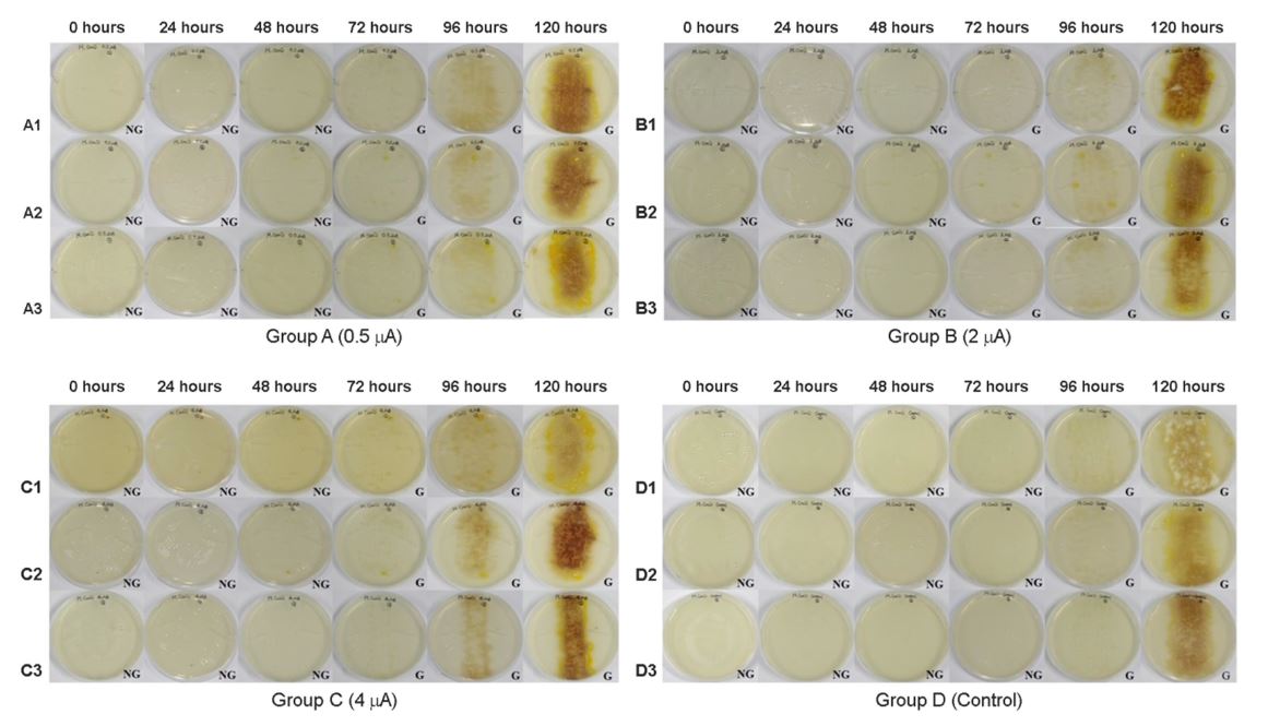
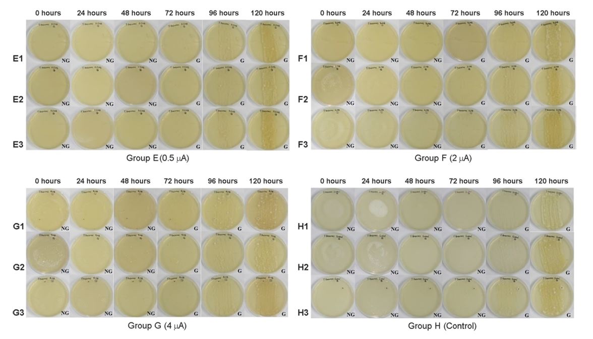
Methods: Inoculums of M. canis and T. tonsurans were applied to twenty-four PDACT (potato dextrose agar-corn meal-Tween 80) plates with a sterile spreader. Petri dishes were allocated into 8 groups according to the fungi species and the amperage delivered to these dishes. Group A, B, C and D were M. canis group and E, F, G, H were T. tonsurans group. The given amperage of electric current was 0.5 μA in group A and E, 2 μA in B and F, 4 μA in C and G. No electric current was given in group D and H.
Results: In groups A, B, and C the average time elapsed for colony appearances were 42 hours, 43.17 hours, and 40.5 hours respectively. The average time elapsed in the control group D was 88.67 hours. In groups E, F, and G the average time elapsed for colony appearances were 63.67 hours, 61.83 hours, and 64.17 hours respectively. The average time elapsed in the control group H was 90.60 hours.
Conclusion: With electric current, faster fungal growth was observed in the amperage range used in this study. Based on these results, we hypothesized that microcurrent helps the fungal growth.
Keywords
Electric stimulus Fungal growth Microcurrent Microsporum canis Trichophyton tonsurans
Dermatophytosis is a commonly encountered condition that is recurrent in its nature, further engendering public health concerns. The optimal method of identifying fungal infection is through culture study, where the specimen is incubated in a specific culture medium. However, this measure is time consuming and defers appropriate time for diagnosis. In respect to this matter, stimulating fungal growth without fungicidal effects has been suggested to affect the growth and reduce the time required for the appropriate diagnosis.
Despite the gravity of clinical impact of fungal infection, the majority of researches in relation to modifying the growth of pathogens are focused on its concurrent antibacterial effects. However, recent studies suggest possible modification of fungal growth through electric current stimulation[1].
A pilot study by Kwon et al.[2] showed faster fungal growth with microcurrent electric stimulation. The involved species in that study was Trichophyton (T.) rubrum, one of the most common fungal pathogen responsible for tinea pedis and onychomycosis.
The primary aim of our study was to confirm the results of the previously described pilot study using the same microcurrent to stimulate the fungal growth. The secondary aim was to evaluate the effects of microcurrent effects on the growth of other fungal species, such as Microsporum (M.) canis and Trichophyton (T.) tonsurans which also are common causes of dermatophytosis[3]-[6].
1. Materials
1) Culture media
All culture media were obtained from Catholic skin clinic, Daegu, Republic of Korea. Nutrient agar was prepared by supplementing potato dextrose agar with corn meal, peptone, Tween 80 (PDACT). To prevent the contamination, we added antibiotics to the media (chloramphenicol 500 mg L-1 and cycloheximide 500 mg L-1).
2) Fungal organisms
Standard sized inoculums of M. canis and T. tonsurans derived from the spore suspension were applied to PDACT plates. The spore suspension was prepared by applying 5 mL of distilled water to a 1-week-old M. canis and 2-week-old T. tonsurans cultures. The inoculums were gently withdrawn by using a sterile pipette and were applied to twenty-four PDACT plates with a sterile spreader. Twenty-four Petri dishes were allocated into eight groups according to the fungi species and the level of amperages delivered to these dishes. Groups A, B, C and D were the M. canis group and E, F, G, H were the T. tonsurans group.
3) Electric device
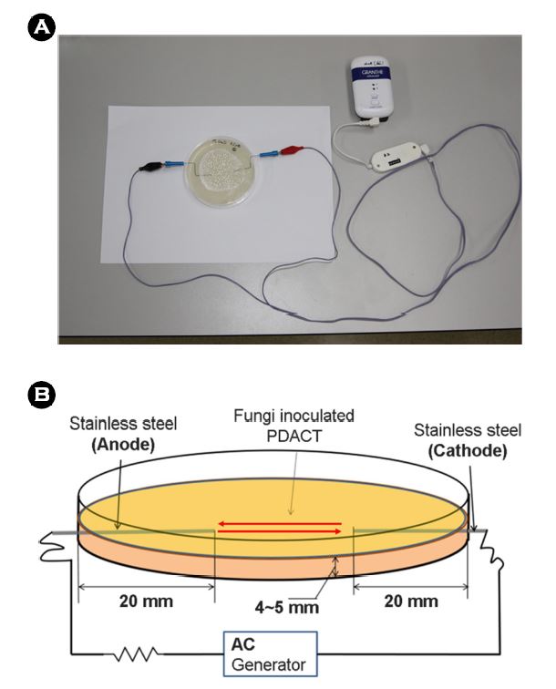
The electric device was operated to generated a series of alternative current of which, the intensity was changeable (9 V, frequency: 8 Hz, Granthe®; Cosmic Co., Seoul, Korea) (Figure 1A, Figure 1B).
2. Methods
All procedures were conducted aseptically to prevent contamination. Two pieces of stainless steel electrodes were used (1 mm in diameter and 2.5 cm long) as cathode and anode electrodes. They were inserted at the top portion of the sterile Petri plate with 2.5 cm apart (Figure 1B). Alternate currents of 0.5 μA, 2 μA, or 4 μA was applied to assigned groups of Petri plates for 30 minutes at room temperature. The given amperage of electric current was 0.5 μA in groups A and E, 2 μA in B and F, 4 μA in C and G respectively. No electric current was given in groups D and H. After electric stimulation, each Petri plate was incubated at 25℃, under natural light with 60~70% of humidity provided by the incubator. The plates were analyzed by interval of every one hour.
Statistical analysis was done using the Statistical Package for the Social Sciences (SPSS) version 19.0 (SPSS, Inc., Chicago, IL, USA) with significance level of < 0.05. Mean and standard deviation is presented in the described data of the paper. Two sample t-test was used in the study for analysis between the experimental and the control groups.
In this study, the experimental groups A, B, C, E, F and G showed faster fungal growth than the control groups D and H, similar with the previous study results by Kwon et al (Figure 2, Figure 3). However there was no significant difference in the level of thickness and onset time of the visible fungal colonies accordingly to the different levels of amperages.
In the M. canis groups of A (0.5 μA), B (2 μA), and C (4 μA) the average time elapsed for colony appearances were 42 hours, 43.17 hours, and 40.5 hours respectively. The average time elapsed in the control group D was 88.67 hours. All groups A, B and C showed statistically significant level of results indicating a shorter time required for colony appearance compared to the control group D (p-value < 0.05). In the T. tonsurans groups of E (0.5 μA), F (2 μA), and G (4 μA) the average time elapsed for colony appearances were 63.67 hours, 61.83 hours, and 64.17 hours respectively. The average time elapsed in the control group H was 90.60 hours. All groups E, F and G showed statistically significant level of results indicating a shorter time required for colony appearance compared to the control group H (p-value < 0.05).
Unlike other reported cases, production of gas bubbles, discoloration, liquefaction, and depression around the electrodes was not observed in this study. The results are summarized in Figure 2 and Figure 3.
In the previous study by Kwon et al.[2], colonies exposed to nano- or microcurrent electric stimulation showed faster fungal seeding than the control groups, which received no electric current. The colonies exposed to the lower level of electric current showed faster fungal growth with denser fungal colonies compared to the colonies exposed to the higher level of electric current. There are, however, some limitations in respect to this finding. First of all, the sample size was small and the study investigation was limited to a single fungal species, T. rubrum. These factors poorly conclude the effect of alternating electric currents on growth of more general population of fungi and its diversity in equal matter. In this study, we sought to determine whether low alternating current could stimulate growth of other fungal species. The results of our study clearly indicate that significantly less incubation time was required for M. canis and T. tonsurans growth after application of microcurrent electric stimulation.
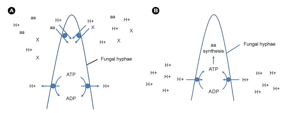
The underlying mechanism of electric stimulation of fungus growth still remains unclear. However, Cheng et al.[7] reported that electric currents could stimulate cell growth. The stimulatory effects of electric current began at 10 μA and inhibitory effects began at 750 μA. In our study, only 0.5 μA to 4 μA levels of electric currents had stimulated the fungal growth. The mechanism of electric current stimulated cellular growth can be proposed in several manners. Adenosine-5'-triphosphate (ATP) generation can play an important role in stimulating cell growth.
When electric current is delivered, the electrons interact with water and produce hydroxyl ions on the cathode side and protons on the anode side of the electrodes. Consequently, a proton gradient is formed between the anode and the cathode interface, leading a shift of proton from anode side to the cathode side within the formed electric field. As the protons reach mitochondrial membrane-bound H+-ATPase, ATP is likely formed[8],[9]. With increased ATP levels, energy required for metabolic activities such as protein synthesis is sufficed and greater amount of proteins could be synthesized (Figure 4A). The increased ATP generation is partially responsible for the increased protein synthesis[10],[11]. Another mechanism of enhanced protein synthesis through electric current stimulation can be achieved by changing the pool of amino acids[12],[13], further increasing the level of amino acid transport (Figure 4B). Thus, the electricity induces ATP generation and increases amino acid availability to cause more protein synthesis and consequently, faster cell growth. An important factor here is that the electric current does not affect DNA metabolism, suggesting that the stimulatory and inhibitory effects of microcurrents on protein synthesis activity occur independently[14].
Our study results indicate the importance of the intensity of microcurrent on stimulating the growth of fungus. On the contrary, Kalinowski et al.[1] suggested that low-voltage direct current electrostimulation acts as a fungicide in a dose-dependent manner. Low-voltage direct current electric stimulation in the range of 500 μA to 3 mA was applied in vivo to T. rubrum. The results of this study clearly demonstrated the fungicidal effect of electric stimulations within the electric current range. The authors proposed that the proposed electric current interfered and caused certain level of damage or even destruction of key cellular enzyme, DNA, cell membranes, or crucial cell transport systems. However, the applied currents used in the study were hundred to a thousand times higher than the electric stimulations used in our study. These high current electric stimulations may generate severe impairment to the cell structures of the fungus. Limitations were exhibited in our study. First of all, the diversity of the fungal species are limited to only M. canis and T. tonsurans. Secondly, the underlying molecular analysis was not conducted and its corresponding molecular mechanism was not derived. Further supporting studies in relation to the effects microcurrent electric stimulation on fungal growth should be carried out with wider ranges of intensity and larger samples sizes with various fungal species to prove more validity to our findings.
In conclusion, electric current retains certain level of stimulatory effects on fungal growth at very low amperages (0.5 μA to 4 μA). Based on the results of our study, microcurrent electric stimulation may be applied to fungal culture and generate potential clinical benefits in respect.
In elaboration, the time elapsed for colonization of fungus could be considerably reduced and provide appropriate manage to the patients in more prompt manner. Additionally, since fungus and its versatile application has been noted in modern manufactured products such as food, beverages, antibiotics and chemical products, electric current stimulation can be utilized for commercial enhancement,
In relation to this article, I declare that there is no conflict of interest.
References
1. Kalinowski DP, Edsberg LE, Hewson RA, Johnson RH, Brogan MS. Low-voltage direct current as a fungicidal agent for treating onychomycosis. J Am Podiatr Med Assoc 2004;94:565-572
Crossref
Google Scholar
PubMed
2. Kwon DR, Kwon H, Lee WR, Park J. Investigating effects of nano- to micro-ampere alternating current stimulation on Trichophyton rubrum growth. Ann Dermatol 2016;28: 575-578
Crossref
Google Scholar
3. Lee WJ, Song CH, Lee SJ, Kim DW, Jun JB, Bang YJ. Decreasing prevalence of Microsporum canis infection in Korea: through analysis of 944 cases (1993-2009) and review of our previous data (1975-1992). Mycopathologia 2012;173:235-239
Crossref
Google Scholar
4. Brajac I, Stojnic-Sosa L, Prpic L, Loncarek K, Gruber F. The epidemiology of Microsporum canis infections in Rijeka area, Croatia. Mycoses 2004;47:222-226
Crossref
Google Scholar
5. Flari ME, Graser Y, Presber W, Tietz HJ. An epidemic of tinea corporis caused by Trichophyton tonsurans among children in Germany. Mycoses 2000;43:191-196
Crossref
Google Scholar
6. Leeming JG, Elliott TS. The emergence of T. tonsurans tinea capitis in Birmingham, U.K. Br J Dermatol 1995; 133:929-931
Crossref
Google Scholar
PubMed
7. Cheng N, Van Hoof H, Bockx E, Hoogmartens MJ, Mulier JC, De Dijcker FJ, et al. The effects of electric currents on ATP generation, protein synthesis, and membrane trans- port of rat skin. Clin Orthop Relat Res 1982;171:264 -272
Crossref
Google Scholar
8. Mitchell P. Chemiosmotic coupling in oxidative and photosynthetic phosphorylation. Biol Rev Camb Philos Soc 1966;41:445-502
Crossref
Google Scholar
PubMed
9. Mitchell P. Vectorial chemistry and the molecular mech- anism of chemiosmotic coupling: Power transmission by proticity. Biochem Soc Trnas 1976;4:399-430
Crossref
Google Scholar
10. Kaziro Y. The role of guanosine-5'-triphosphate in poly- peptide chain elongation. Biochim Biophys Acta 1978; 505:95-127
Crossref
Google Scholar
PubMed
11. Keller EB, Zamecnik PC. The effects of guanosine diphos- phate and triphosphate on the incorporation of labeled amino acids into proteins. J Biol Chem 1956;221:45-59
Crossref
Google Scholar
PubMed
12. Hubel KA. The effects of electrical field stimulation and tetrodotoxin on ion transport by isolated rabbit ileum. J Clin Invest 1978;62:1039-1047
Crossref
Google Scholar
PubMed
13. Gow NAR. Relationship between growth and the electrical current of fungal hyphae. Biol Bull 1989;176:31-35
Crossref
Google Scholar
PubMed
14. Bozoky L, Kiszely G, Hoffman TA, Ladik J. Effect of electro- static fields on cell mitosis. Nature 1963;199:1306
Crossref
Google Scholar
PubMed
Congratulatory MessageClick here!
