pISSN : 3058-423X eISSN: 3058-4302
Open Access, Peer-reviewed
pISSN : 3058-423X eISSN: 3058-4302
Open Access, Peer-reviewed

Jun Suk Hong,Dong Won Lee,Moo Kyu Suh,Gyoung Yim Ha
10.17966/JMI.2018.23.4.118 Epub 2019 January 02
Abstract
Keywords
Morphology Trichophyton verrucosum
Trichophyton verrucosum, a zoophilic dermatophyte, is distributed worldwide and is the most common causative pathogen of dermatophytosis caused by cattle1,2. T. verrucosum is occasionally transmitted to humans by direct contact with an infected cattle's skin. Since Kim et al. reported the first case of dermatophytosis caused by T. verrucosum isolated from a cattle in Korea in 1986, 10 more cases of dermatophytoses caused by cattle have been reported thereafter. The common clinical manifestations of T. verrucosum are sycosis and tinea corporis, usually on exposed skin surfaces. Because infections due to T. verrucosum are usually characterized clinically by intense inflammation, they are often misdiagnosed as eczema, herpes, or bacterial folliculitis3.
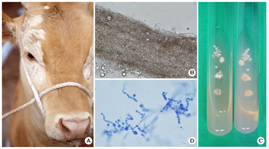
The infection presents with multiple, walnut to child-palm-sized, whitish plaques on the scalp and face of cattle infected with T. verrucosum (Figure 1A). Direct microscopic examination of a hair infected with T. verrucosum reveals several chains of chlamydoconidia present around the hair shaft (Figure 1B). The macroscopic morphology of T. verrucosum reveals that it is slow-growing, folded, heaped, and glabrous; they form white colonies, lacked pigment on the reverse side (Figure 1C), and the microscopic morphology of T. verrucosum shows chains of chlamydoconidia (Figure 1D).
The characteristic microconidia are tear-shaped microconidia and, less frequently, string-bean-shaped, and 3-5-celled macroconidia on blood agar enriched with thiamine and inositol1. Microscopic examination and macroscopic morphology are necessary for the identification of causative organisms; however, physicians can identify species more precisely by molecular biological analyses.
In relation to this article, We declare that there is no conflict of interest.
References
1. Kwon-Chung KJ, Bennett JE. Medical mycology. Philadelphia: Lea & Febiger, 1992:148-149
Crossref
2. Chermette R, Ferreiro L, Guillot J. Dermatophytoses in animals. Mycopathologia 2008;166:385-405
Crossref
Google Scholar
PubMed
3. Romano C, Massai L, Gianni C, Crosti C. Case reports. Six cases of infection due to Trichophyton verrucosum. Mycoses 2001;44:334-337
Crossref
Google Scholar
PubMed
Congratulatory MessageClick here!
