pISSN : 3058-423X eISSN: 3058-4302
Open Access, Peer-reviewed

pISSN : 3058-423X eISSN: 3058-4302
Open Access, Peer-reviewed

Hye-Jin Ahn,Kyung-Gu Chung,Min Kyung Shin
10.17966/JMI.2026.31.3.118 Epub 2026 October 01
Abstract
The spectrum of conditions that lead to an immunocompromised state in patients is broad. Primary immunodeficiencies often result from genetic mutations. Acquired immune dysfunction may result from infection-mediated immune deficiency, underlying diseases, and medications. The immunocompromised patient may manifest a variety of cutaneous conditions that can be challenging to diagnose and manage. Among cutaneous manifestations, the major category seen in immunocompromised patients is infections. Immunocompromised patients may exhibit either unusually severe or prolonged skin infections with common pathogens or infections with opportunistic organisms. Cell-mediated immunity is driven by CD8+ cytotoxic T-cells via antigens presented on MHC class I and macrophages activated by Th1 cells. It defends against intracellular pathogens, including mycobacteria, filamentous bacteria, dimorphic or opportunistic fungi, and some protozoa through apoptosis and cell lysis. Impaired cell-mediated immunity also predisposes to reactivation of latent viruses. Neutropenia is defined as a decrease in the absolute neutrophil count (ANC) below 1,500/μL. Skin flora can be pathogenic and cause bacterial sepsis in neutropenic patients. Hospitalized neutropenic patients have a higher risk of developing systemic infection due to opportunistic angioinvasive fungi. Primary and secondary humoral immunodeficiencies can affect B cell production and function, antibody formation and function, and the effectiveness of the complement pathway. Deficiency in any of these components can put patients at particular risk of infection with encapsulated bacteria by impairing opsonization for phagocytosis. This review focuses on opportunistic cutaneous infections occurring in immunocompromised patients.
Keywords
Cellular immunity Humoral immunity Immunocompromised host Neutropenia Opportunistic infections
Immunocompromised patients develop either unusually severe or prolonged skin infections with common pathogens or opportunistic organisms. A wide spectrum of conditions may contribute to the development of an immunocom- promised state, ranging from primary immunodeficiencies (caused by genetic mutations) to acquired immune dysfunc- tion resulting from infection-mediated immune deficiency (e.g., acquired immune deficiency syndrome [AIDS]), comorbidities (e.g., malignancies and diabetes mellitus), and medications (e.g., chemotherapy and biologic agents)1. This review offers a practical, clinically oriented approach for characterizing rare opportunistic cutaneous infections in immunocompro- mised patients, with special emphasis on epidemiological and diagnostic considerations relevant to South Korea. These infections may be rare or potentially fatal and may present with distinctive cutaneous manifestations or remain relatively undetected by dermatologists.
Physiologically, cell-mediated immunity (CMI) defends against intracellular pathogens through apoptosis and cell lysis2. It is driven by CD8+ cytotoxic T-cells via antigens pre- sented on the major histocompatibility complex class I and macrophages activated by type 1 T helper (Th1) cells. As a result, immunocompromised patients are susceptible to opportunistic infections and are at an increased risk of latent viral reactivation.
2.1 Cutaneous tuberculosis (TB)
Cutaneous TB is typically acquired through exogenous inoculation, contiguous spread, or hematogenous spread3. One such entity is TB cutis orificialis, which arises from auto- inoculation of mucosal surfaces or spread from advanced pulmonary, gastrointestinal, or genitourinary TB, and occurs in patients with low immunity. It usually appears as one or more ulcerative, painful lesions in the oral, genital, or anal mucosa and periorificial skin3,4. Miliary or disseminated TB is another life-threatening form of TB that occurs primarily in children and infants following an infection, such as measles or scarlet fever, that diminishes their immune response. Another example is TB re-emergence in patients infected with human immunodeficiency virus (HIV) and having a CD4 count <100 cells/μL.
Cutaneous TB in immunocompromised patients is char- acterized by a multibacillary form in which the lesions are often closely packed and teeming with mycobacteria (acid-fast bacilli [AFB])3. Given that diminished CMI may obscure the development of characteristic histopathological features, such as caseating granulomas, AFB staining should be per- formed when mycobacterial infection is suspected5. However, the results of the tuberculin test and interferon-gamma release assay may be negative or indeterminate in immunocompro- mised patients due to impaired host defense mechanisms4,6. The management of cutaneous TB follows the same treat- ment principles as systemic TB.
2.2 Non-tuberculous mycobacteria
Non-tuberculous mycobacteria (NTM) can be found in soil, water, and almost anywhere in the world. These infections predominantly occur in immunocompromised patients or after skin trauma in immunocompetent patients3. NTM infections due to rapidly growing mycobacterial (RGM) species (in- cluding M. fortuitum, M. abscessus, and M. chelonae) are associated with skin trauma and injection sites. In particular, M. chelonae and M. abscessus infections present as dissem- inated cutaneous infections in immunocompromised patients3. It has been noted that patients with RGM infections with multiple lesions are significantly more likely to be receiving immunosuppressive therapy than those presenting with a single lesion7. In immunocompromised patients, cutaneous infections caused by slowly growing mycobacteria, such as M. kansasii or the M. avium complex, usually occur after hematologic dissemination from pulmonary lesions8,9. Since NTM is resistant to most anti-tuberculous drugs, the treatment is often long and difficult3. In immunocompetent patients, therapy consists of single or dual therapy with drugs such as clarithromycin, minocycline, doxycycline, or trimethoprim-sulfamethoxazole (TMP-SMX), or combination therapy with ethambutol and rifampicin3. Immunocompromised patients are treated for a longer duration and may require long-term suppressive therapy2.
2.3 Filamentous bacteria
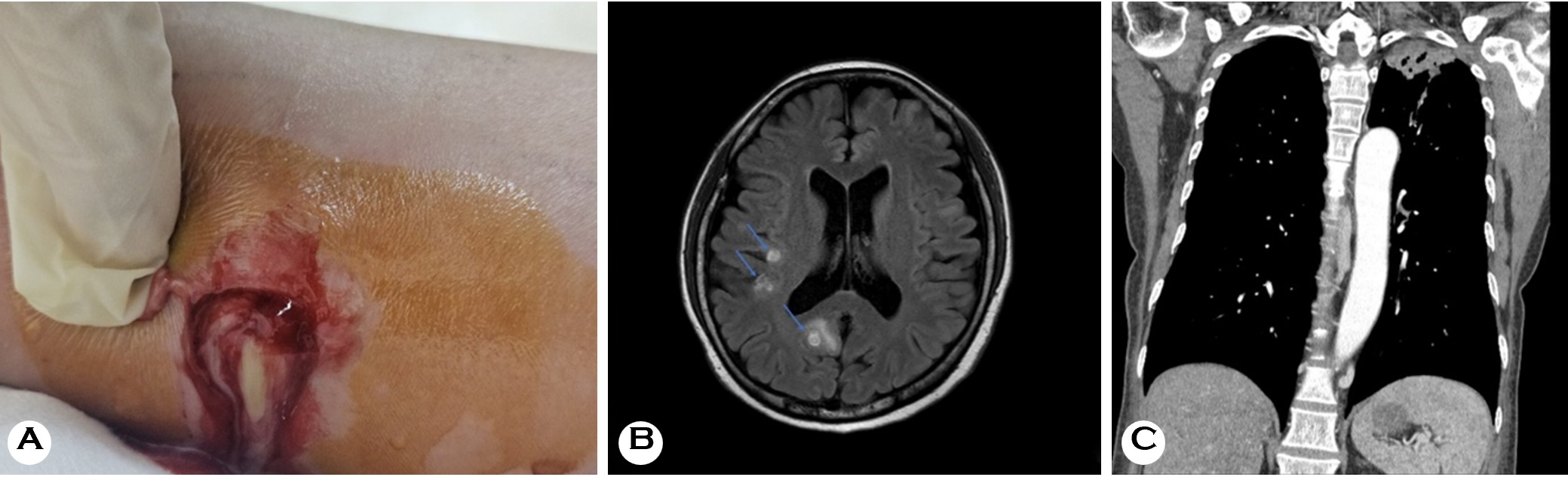
Actinomycetoma is caused by Gram-positive, branching, filamentous bacteria, most commonly Nocardia and Actino- madura species. In contrast, Actinomyces, a commensal flora of the oropharynx, gastrointestinal tract, and urogenital tract, is an uncommon cause of actinomycetoma. Patients with esophageal actinomycosis are usually immunosuppressed due to malignancy, HIV, or solid organ transplant10. Nocardia is a common environmental pathogen. In recent years, the incidence of Nocardia infection cases and detections has increased significantly, presumably because of an increase in the number of immunosuppressed people, such as those receiving organ transplants or undergoing cancer chemo- therapy11. Furthermore, immunocompromised status was associated with an increased risk of disseminated infection (Fig. 1) and poor outcomes11. Actinomycosis and nocardiosis have several distinct clinical manifestations, ranging from superficial skin and soft tissue infection to chronic mycetoma form. Furthermore, actinomycetomas should be differen- tiated from eumycetomas, which are deep fungal infections2. Patients with actinomycosis require prolonged (6-12 months) high-dose penicillin G or amoxicillin therapy10; however, in case of actinomycetoma or Nocardia infection, TMP-SMX and amikacin are used in combination, and surgical excision is performed if necessary2.
2.4 Fungi
2.4.1 Dimorphic fungi
Common dimorphic fungi, including Blastomyces, Cocci- dioides, Histoplasma, Talaromyces, and Emergomyces species, are found in specific geographical environments, existing as molds and transforming into pathogenic yeasts (or spherules) at human body temperature. The lungs are the most common site of endemic mycoses, with disease course ranging from subclinical respiratory infection to life-threatening systemic disease2. Under healthy conditions, T-cells release interferon-gamma (IFN-γ) and tumor necrosis factor-alpha (TNF-α), which activate macrophages to destroy the engulfed fungi. In hosts with compromised CMI, this cytokine signaling is lacking or blunted, allowing the fungus to replicate unchecked and disseminate via the bloodstream12.
Histoplasmosis is caused by Histoplasma capsulatum var. capsulatum and is extremely rare in South Korea. Lungs are the most common site of primary infection, with 11% of patients with AIDS developing skin manifestations13. Histo- plasmosis tends to disseminate, resulting in papular lesions on the face and the trunk2. Talaromycosis, caused by Talaromyces marneffei, is another opportunistic infection seen in indi- viduals with advanced HIV infection in endemic tropical and subtropical regions of Southeast Asia and southern China. It often presents with papular skin lesions with central necrosis or umbilication14.
2.4.2 Encapsulated yeasts
Cryptococcosis, caused by Cryptococcus neoformans and Cryptococcus gattii, presents mainly as meningoencephalitis; however, the disease may range from asymptomatic lung colonization to systemic dissemination. The unique fungal polysaccharide capsule of Cryptococcus suppresses the innate immune recognition of the fungus; therefore, a strong adap- tive immune response, including CD4+ helper T-cell activity, is required for fungal control. Cutaneous cryptococcosis typically occurs in patients with advanced HIV/AIDS13, presenting as subcutaneous papules and nodules on the face and neck. It should be suspected in patients presenting with papulonod- ular necrotizing lesions with central umbilication resembling molluscum contagiosum2.
2.4.3 Candida
Candida species are opportunistic pathogens capable of causing a variety of mucocutaneous and systemic diseases in immunocompromised hosts. Chronic mucocutaneous candi- diasis comprises a heterogeneous group of syndromes char- acterized by recurrent or chronic noninvasive symptomatic infections of the skin, nails, and mucous membranes, usually caused by C. albicans15. Notably, oral candidiasis is a marker for an increased rate of progression to AIDS16, and the pre- sence of oral candidiasis in AIDS may be associated with poor survival in some patients17. Cutaneous candidiasis often involves intertriginous skin, with some patients developing markedly thickened areas with gross hyperkeratosis18. Par- onychia is a common clinical manifestation of cutaneous candidiasis often accompanied by extensive nail plate involve- ment and total dystrophic onychomycosis18,19. Various host factors that increase susceptibility to superficial infections due to Candida species include autosomal dominant hyper-IgE syndrome, gain-of-function (GOF) STAT1 defects, CARD9 defects, and autoimmune polyendocrinopathy-candidiasis-ectodermal dystrophy syndrome15. In contrast, invasive candi- diasis occurs predominantly in the setting of a clear breach in host defense, risk factors for which include substantial disruption of the mucosal barrier (e.g., chemotherapy-induced mucositis) combined with underlying neutropenia20. C. albicans is the predominant cause of hematogenous Candida infections and invasive candidiasis worldwide, although the incidence of infection with non-albicans species is increas- ing20,21. A systematic review of 100 documented cases of disseminated candidiasis with skin involvement found that 68% of cases were caused by C. tropicalis, followed by C. krusei (15%)22. The systematic review also noted that the most frequent underlying disease was acute leukemia, and the most frequent pattern of skin lesions was erythematous or purpuric maculopapular lesions (44%), followed by purpuric or erythematous papular lesions with a pale center (22%), nodules (17%), and subcutaneous abscess (6%)22.
2.5 Protozoa
Leishmaniasis is a zoonotic protozoan parasitic disease transmitted by sandflies infected with species of Leishmania. Although an estimated 600,000-1,000,000 new cases of cutaneous leishmaniasis (CL) and 50,000-90,000 new cases of visceral leishmaniasis (VL) occur worldwide each year23, it is extremely rare in South Korea. In immunocompetent in- dividuals, it most commonly manifests as CL, which presents as plaques with indurated borders and central ulceration that may heal spontaneously with atrophic scarring2. However, VL is an opportunistic infection in persons with HIV/AIDS or other causes of CMI suppression24. Infected individuals typically develop a strong Th1-mediated immune response25; however, if CMI is impaired, parasites may remain dormant in scars or lymph nodes after treatment and reactivate24. Giemsa-stained skin scrapings typically show many intracellular amastigotes inside a mononuclear cell cytoplasm25. Lately, more sensitive molecular diagnostic methods, particularly polymerase chain reaction (PCR), are being used to confirm the diagnosis of leishmaniasis2. There is no universally accepted first-line treatment of CL; the available systemic therapies are associated with substantial toxicities and show variable efficacy across different Leishmania species. Because the host's CMI plays a critical role in controlling the infection, treatment for CL should be tailored according to the patient, the infecting Leishmania species, and the region in which the infection was acquired26.
2.6 Latent virus
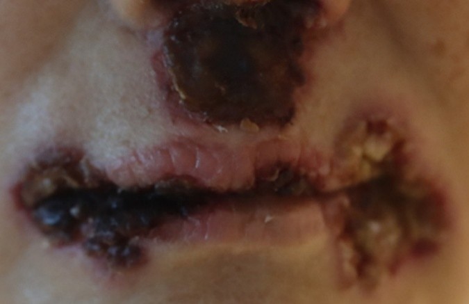
Among viral pathogens, recurrent human herpes simplex virus (HSV) infection is common in immunocompromised patients. Chronic ulcerative perianal disease caused by HSV-2 was one of the first features of AIDS to be reported27. Immunocompromised patients may develop further severe manifestations of HSV infection (Fig. 2). Additionally, refrac- tory or multi-dermatomal varicella-zoster virus (VZV) infection can be seen with iatrogenic immunosuppression. A study reported that the increased risk of VZV reactivation observed with Janus kinase inhibitors may be attributable to their suppression of type I and II interferon signaling, which plays a critical role in maintaining VZV latency and preventing viral reactivation (Fig. 3)28.

Cytomegalovirus (CMV) infections are generally asymptom- atic or mild in immunocompetent individuals. CMV remains dormant over the life of the host in myeloid cells, reactivating in the setting of immunosuppression29. Cutaneous CMV infection is rare but may indicate disseminated disease and an unfavorable prognosis. A clinicopathologic series including older patients reported approximately 85% mortality within six months30. A recent case series described cutaneous ulcers caused by CMV in patients with malignancy and organ transplantation; three of the four patients died soon after diagnosis of CMV infection despite ganciclovir treatment29. Ulceration is the most common presentation of CMV infec- tion; other cutaneous findings include necrotizing vasculitis, thrombosis leading to limb ischemia, and drug reaction with eosinophilia and systemic symptoms (DRESS)/drug-induced hypersensitivity syndrome29.
Neutrophils are the primary mediators of rapid innate host defense against most bacterial and fungal pathogens that activate before the complex humoral and lymphocyte cellular processes of acquired immunity act on an infection31. Thus, neutropenic patients are particularly susceptible to bacterial and angioinvasive fungal infections. Neutropenia is defined as a decrease in the absolute neutrophil count (ANC) below 1,500/μL. Patients can develop neutropenia due to decreased production in the bone marrow or increased peripheral destruction. It can arise in childhood from congenital factors or in adulthood through acquired factors32. The risk of infec- tion increases with the severity and duration of neutropenia, particularly in patients with severe (ANC < 500/μL), profound (ANC < 100/μL), or prolonged (>7 days) neutropenia33.
3.1 Invasive bacterial infections
Common pathogens that cause bacterial sepsis in neutro- penic patients include both Gram-negative and Gram-positive bacteria, such as Staphylococcus aureus, Pseudomonas aeruginosa, and Klebsiella pneumoniae, which can be acquired from skin disruption due to an intravenous catheter, proced- ures, or surgeries32. Lately, hospital- and community-acquired Stenotrophomonas maltophilia infections are being reported as an emerging opportunistic pathogen34-36. It is an aerobic Gram-negative ubiquitous bacillus with low virulence and is considered an uncommon pathogen in immunocompetent individuals; however, in immunosuppressed hosts, especially neutropenic individuals, on chemotherapy or broad-spectrum antibiotics, S. maltophilia can cause neutropenic septicemia34.
3.2 Angioinvasive fungi
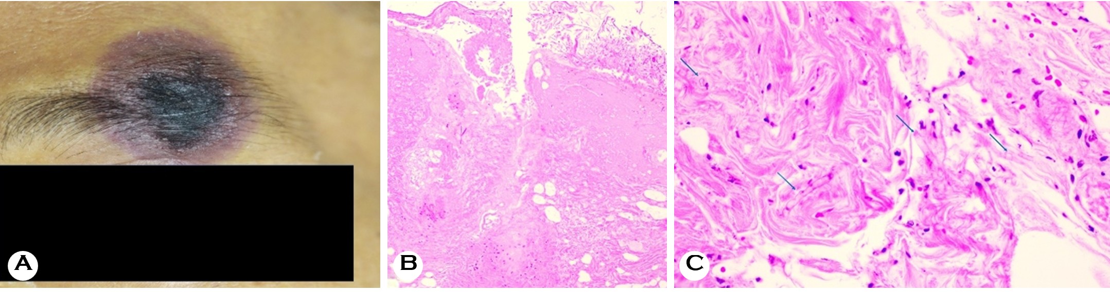
Hospitalized neutropenic patients are also at a greater risk of contracting angioinvasive fungal infections from Aspergillus, Mucor, Rhizopus, and Fusarium32. Filamentous hyphal struc- tures are too large for macrophages to engulf; thus, neutro- phils become the key defenders against fungi by releasing reactive oxygen species and antifungal enzymes from their granules and forming neutrophil extracellular traps that capture and damage fungi37. Neutropenic patients lack these defense mechanisms, making them particularly susceptible to opportunistic angioinvasive fungal infections that can in- vade blood vessel walls, resulting in fungemia and significant morbidity and mortality. Primary cutaneous fungal infections can occur at sites of percutaneous damage from an intra- venous catheter or via respiratory or gut inoculation32,38. Other sources of primary skin infections include onychomycosis caused by Aspergillus or Fusarium39-41. Skin involvement is the first clue in most of the disseminated fusariosis cases and often occurs at an early stage of the disease. A study reported that among 16 patients with fusariosis with dis- seminated skin lesions, 11 had a recent history of Fusarium cellulitis at the site of onychomycosis, 3 had local trauma, and 2 had an insect bite, corroborating that skin was the primary site of infection41. Clinical manifestations of angioinvasive fungal infection include multiple painful erythematous macular or papular lesions, which usually have a necrotic center (Fig. 4)41,42. Notably, fungal infections can be mistaken for bacterial infections. In immunocompromised patients, clinicians should consider the diagnosis of mucormycosis in patients pres- enting with necrotic sinusitis or paranasal cellulitis, as well as Fusarium infection presenting with ecthyma gangrenosum-like lesions that may be mistaken for pseudomonal ecthyma42.
While antibody deficiencies can also occur as primary dis- orders due to inherited genetic defects, secondary immune disorders are far more prevalent and can be caused by various diseases and their treatment, certain medications, and surgical procedures32,43. Both primary and secondary humoral immunodeficiencies can affect B-cell production and function, antibody formation and function, and the effect- iveness of the complement pathway43. Deficiency in any of these components can put patients at particular risk of infection from encapsulated bacteria, including Streptococcus pneumoniae, Haemophilus influenzae, Streptococcus aga- lactiae, and Neisseria species, due to the impairment of opsonization for phagocytosis and lysis of pathogens32,44.
Deficiencies of the terminal complement components (C5-C9) confer a particularly high susceptibility to invasive Neisseria infections45. Functional or anatomic asplenia hampers the clearance of opsonized encapsulated bacteria from the blood- stream, increasing the severity and risk of infections caused by encapsulated organisms44.
4.1 Meningococcemia
Meningococcemia is caused by Neisseria meningitidis. Cutaneous manifestations of meningococcemia may aid in early diagnosis, which is crucial to prevent disease progression. Acute cutaneous manifestations result from disseminated intravascular coagulation (DIC), inducing thrombocytopenia and widespread thrombosis; this type of hemorrhagic rash is more frequently observed in fatal cases46. Purpura fulminans is a rapidly evolving syndrome of skin microvessel thrombosis and hemorrhagic necrosis and a severe complication of invasive N. meningitidis infection with a high mortality rate47. Congenital terminal complement deficiency increases the relative risk of meningococcal infection by 1,000-fold com- pared to the general population48. Similarly, complement inhibitor therapy (eculizumab/ravulizumab) renders patients highly susceptible to this infection49. Similarly, lose opsoni- zation and IgM-mediated early defense against encapsulated organisms (S. pneumoniae, N. meningitidis, and H. influ- enzae), placing them at lifelong risk of overwhelming post-splenectomy infection and its accompanying purpura fulmi- nans (DIC-mediated skin necrosis)50.
Cutaneous lesions in immunocompromised patients with disseminated opportunistic infections must be considered an important clue to an underlying life-threatening condition. Since the causative pathogen is often difficult to determine on the basis of clinical manifestations alone, an incisional skin biopsy and microbial study can be useful. Serum pro- calcitonin levels may also help distinguish bacterial infection from other causes, whereas serum fungal markers, such as beta-D-glucan, may facilitate the diagnosis of invasive fungal disease18. A summary of representative cutaneous infections stratified according to the underlying immune defect, along with their characteristic cutaneous clues and diagnostic approaches, is given in Table 1. Clinicians should be cautious while evaluating cutaneous lesions in immunocompromised patients because of the broad range of potential microbial etiologies and the unusual clinical manifestations of common infections in this population.
|
Immune defect |
Representative |
Characteristic |
Diagnostic |
|
Cellular |
Mycobacterium |
Chronic plaques, nodules,
or ulcers; periorificial ulceration; purpuric or umbilicated papules in
disseminated TB |
Skin biopsy for
histopathology, AFB staining, and mycobacterial culture ± PCR |
|
Non-tuberculous |
Papules, nodules,
abscesses, ulcers, |
Skin biopsy for
histopathology, AFB staining, and mycobacterial culture ± PCR |
|
|
Actinomyces spp. / |
Indurated nodules or
abscesses with |
Tissue biopsy with
appropriate stains and bacterial culture |
|
|
Cryptococcus |
Molluscum-like or
umbilicated papules, |
Skin biopsy for
histopathology and fungal culture |
|
|
Talaromyces |
Papules with central
necrosis or umbilication |
Skin biopsy for
histopathology and fungal culture |
|
|
Leishmania spp. |
Plaques with indurated
borders and |
Skin biopsy for
histopathology, |
|
|
Candida spp. |
Chronic mucocutaneous candidiasis; |
Fungal culture and skin
biopsy |
|
|
HSV / VZV |
Chronic or atypical ulcers
(HSV); |
PCR of the lesional
material |
|
|
CMV |
Chronic, deep, or necrotic
ulcers, |
Skin biopsy for
histopathology and |
|
|
Neutropenia |
Pseudomonas |
Ecthyma gangrenosum:
hemorrhagic |
Blood culture; skin biopsy
for histopathology and bacterial |
|
Candida spp. |
Invasive candidiasis:
erythematous or |
Blood culture; skin biopsy
for histopathology and fungal |
|
|
Aspergillus spp. |
Papules, nodules, or
plaques with hemorrhage or central necrosis; |
Skin biopsy for
histopathology |
|
|
Fusarium spp. |
Multiple purpuric macules,
papules, or |
Blood culture; skin biopsy
for histopathology and fungal culture |
|
|
Mucorales |
Rapidly progressive
necrotic plaques or |
Skin biopsy for
histopathology |
|
|
Humoral/complement |
Neisseria |
Petechiae, purpura,
ecchymoses; |
Blood culture and PCR |
|
Abbreviations: AFB, Acid-Fast Bacillus; CMV, Cytomegalovirus; HSV,
Herpes Simplex Virus; PCR, Polymerase Chain Reaction; TB, Tuberculosis; VZV, Varicella-Zoster Virus |
|||
References
1. 1. Hoeger PH, Kinsler V, Yan AC, Harper J, Oranje AP, Bodemer C, et al. Harper's Textbook of Pediatric Dermatology. 4th ed. Hoboken: John Wiley & Sons; 2019
Congratulatory MessageClick here!