pISSN : 3058-423X eISSN: 3058-4302
Open Access, Peer-reviewed
pISSN : 3058-423X eISSN: 3058-4302
Open Access, Peer-reviewed

Jisu Kim,Seung Hee Cho,In Young Yoo,Ji Hyun Lee
10.17966/JMI.2026.31.2.88 Epub 2026 June 30
Abstract
Keywords
Dermatophytosis Microsporum canis Tinea capitis
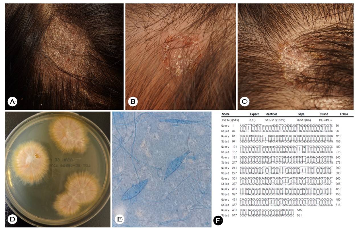
A 7-year-old girl presented with a 1-week history of patchy alopecia on the scalp. She had been in close contact with cats during her travel 6 weeks earlier. Examination revealed three scaly, erythematous patches on the frontal, vertex, and occipital scalp (Fig. 1A-C). Further examination using potassium hydroxide (KOH) microscopy showed septate hyphae with ectothrix spores that surrounded the hair shafts. Fungal culture on Sabouraud's dextrose agar produced whitish-yellow cotton-like colonies at 7 days (Fig. 1D), with macroconidia that exhibited variably thick echinulate walls of 5-15 cells with curved knob-like ends (Fig. 1E). Polymerase chain reaction (PCR) identified Microsporum canis within 4 days (Fig. 1F). The patient was given an 8-week treatment with oral itraconazole (100 mg/day; i.e., 4.1 mg/kg/day) and topical isoconazole, resulting in clinical improvement.
Tinea capitis predominantly affects children, and it is clinically significant due to the risk of permanent hair loss in the case of delayed diagnosis1. This condition is most com- monly caused by species of Trichophyton and Microsporum; Microsporum canis, transmitted by animals such as cats and dogs, is a predominant cause in Korea2. Diagnosis of the condition is primarily based on clinical findings, supported by KOH microscopy and fungal culture, where the latter allows for definitive species identification. Recent evidence indicates that dermatophyte PCR is a rapid and highly sensitive tool for the baseline diagnosis of pediatric tinea capitis, enabling early species-level identification and timely initiation of appropriate systemic therapy3.
This case demonstrates that a comprehensive diagnostic approach using microscopy, fungal culture, and PCR enables early and accurate species-level diagnosis, thereby preventing complications such as scarring alopecia.
References
1. Leung AKC, Hon KL, Leong KF, Barankin B, Lam JM.Tinea capitis: An updated review. Recent Pat Inflamm Allergy Drug Discov 2020;14:58-68
Google Scholar
2. Park SK, Park SW, Yun SK, Kim HU, Park J. Tinea capitis in adults: A 18-year retrospective, single-centre study in Korea. Mycoses 2019;62:609-616
Google Scholar
3. Theiler M, Luchsinger I, Rast AC, Schwieger-Briel A, Weibel L, Bosshard PP. Precision diagnostics in paediatric dermatology: Advancing management of tinea capitis through dermatophyte PCR. J Eur Acad Dermatol Venereol 2025;39:398-403
Google Scholar
Congratulatory MessageClick here!
