pISSN : 3058-423X eISSN: 3058-4302
Open Access, Peer-reviewed
pISSN : 3058-423X eISSN: 3058-4302
Open Access, Peer-reviewed

Halim Jo,Ji Hyun Lee
10.17966/JMI.2026.31.2.86 Epub 2026 June 30
Abstract
Keywords
Cosmetic techniques Mycobacterium abscessus Nontuberculous mycobacterial infection
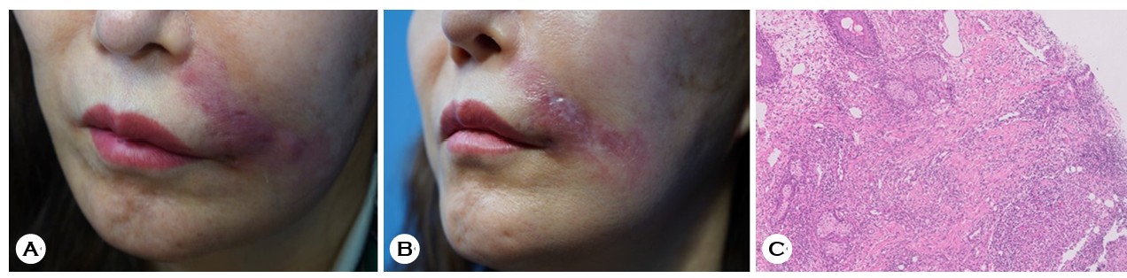
A 60-year-old woman with a history of hypothyroidism presented with a 1-month history of a solitary, indurated, erythematous-to-violaceous plaque on the left perioral area following an unspecified cosmetic injection performed by a non-dermatologist (Fig. 1). She reported daily manipulation (squeezing) of the lesion. Prior antimicrobial therapy at another hospital had been ineffective. She denied fever or other systemic symptoms. Routine laboratory investigations, including complete blood count, erythrocyte sedimentation rate, and C-reactive protein, were within normal limits. Anti- nuclear antibody was positive (titer 1:80, homogeneous). Histopathological examination demonstrated chronic inflam- matory cell infiltration without granuloma formation (Fig. 1). Mycobacterial culture yielded Mycobacterium abscessus (M. abscessus), and species identification was subsequently confirmed by polymerase chain reaction. Empiric ciprofloxacin and clarithromycin were initiated and later adjusted based on antimicrobial susceptibility testing. However, the lesion continued to enlarge with increased induration and inter- mittent oozing over the subsequent 3 months, after which the patient was lost to follow-up.
M. abscessus is an emerging cause of cutaneous non- tuberculous mycobacterial (NTM) infection, typically presenting as erythema, indurated plaques, nodules, or abscesses1-3. This rapidly growing mycobacterium is widely found in soil and water and may persist despite exposure to standard disinfectants, thereby contaminating procedural instruments, tap water, and injectable materials2. With the increasing global use of cosmetic procedures, particularly those performed by non-dermatologists, procedure-associated M. abscessus in- fections have been increasingly reported. Reported sources include hyaluronic acid filler and botulinum toxin injections, lipolysis injections, microdermabrasion, mesotherapy and acupuncture with thread embedding2-4.
Although NTM infections are generally regarded as oppor- tunistic infections in immunocompromised hosts, most procedure-associated cases occur in immunocompetent individuals, making early recognition challenging4. In a retro- spective study, the mean incubation period of NTM was 3 weeks and the mean time to diagnosis was 9.8 weeks3. Cutaneous lesions typically follow a delayed and indolent course, frequently leading to misdiagnosis as bacterial infection or a foreign-body granuloma2,3. Therefore, mycobacterial culture with molecular confirmation should be pursued when the clinical course is suggestive.
Treatment of M. abscessus infections remains particularly challenging because of its intrinsic resistance to multiple antibiotics1. Although macrolides remain the therapeutic backbone, combination regimens with one or two additional agents are generally recommended, and definitive treatment should be guided by susceptibility testing3,4.
This case highlights the importance of considering M. abscessus infection in patients presenting with persistent inflammatory lesions following cosmetic procedures. Lesions that fail to respond to conventional antimicrobial therapy should raise suspicion for NTM infection. A thorough history of potential exposures, including aesthetic procedures, is essential, as these may represent sources of contamination. Early mycobacterial culture, molecular identification, and susceptibility-guided therapy are critical for improving clinical outcomes and preventing prolonged disease courses.
References
1. Griffith DE, Aksamit T, Brown-Elliott BA, Catanzaro A, Daley C, Gordin F, et al. An official ATS/IDSA statement: Diagnosis, treatment, and prevention of nontuberculous mycobacterial diseases. Am J Respir Crit Care Med 2007; 175:367-416
Google Scholar
2. Sarkar R, Thekho AJ, Rini T. Infectious complications in esthetic dermatology procedures: A narrative review. J Cutan Aesthet Surg. Online ahead of print. doi:10.25259/JCAS_198_2025
3. Zeng R, Xiong J, Gao W, Peng J, Shi Y, Zhang W, et al. Cutaneous nontuberculous mycobacteria infections fol- lowing cosmetic procedures: A retrospective study. Infect Drug Resist 2025;18:2301-2309
Google Scholar
4. Moreno-Izquierdo C, Zurita J, Contreras-Yametti FI, Jara-Palacios MA. Mycobacterium abscessus subspecies abscessus infection associated with cosmetic surgical procedures: Case series. IDCases 2020;22:e00992
Google Scholar
Congratulatory MessageClick here!
