pISSN : 3058-423X eISSN: 3058-4302
Open Access, Peer-reviewed
pISSN : 3058-423X eISSN: 3058-4302
Open Access, Peer-reviewed

Yong Woo Choi,Nuri Na,Yong Joon Bang,Joonsoo Park
10.17966/JMI.2018.23.3.82 Epub 2018 September 29
Abstract
Keywords
Dermatophyte Epidermophyton floccosum Macroconidia
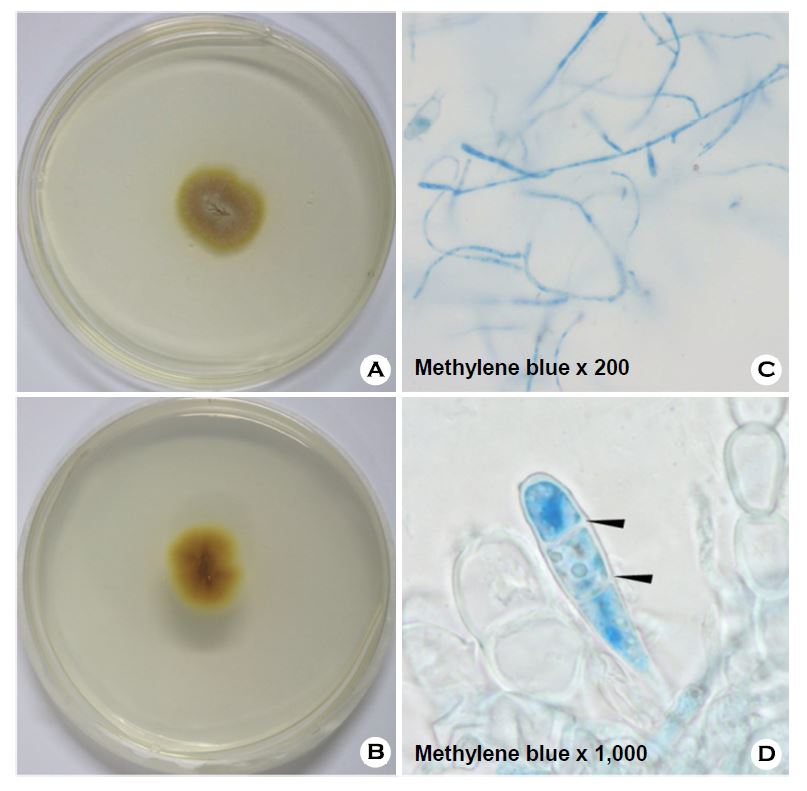
Epidermophyton floccosum is a filamentous fungus that causes skin and nail infections in humans1. This anthropophilic dermatophyte can lead to diseases such as tinea pedis, tinea cruris, tinea corporis, and onychomycosis2. E. floccosum is the only species in the genus Epidermophyton. During the twentieth century, this species was the fourth most common cause of dermatophytosis in North America3. Although this ascomycete has a worldwide distribution, it is more commonly isolated in patients from tropical and subtropical areas2. The non-soil-associated fungus requires no specific growth conditions and displays characteristic smooth club-shaped macroconidia under the microscope2. Diagnostic approaches to the fungal infection include a physical examination, followed by testing skin and other cultures, and molecular detection.
As observed under a microscope, E. floccosum forms dull green or khaki colonies with a suede-like powdery surface that is raised and folded in the center with a flat periphery2. An ocher, deep-yellowish brown to mustard-yellow reverse pigment is usually present2. The morphology of E. floccosum is characteristic of smooth, thin-walled macroconidia, which are often produced in clusters and grow directly from hyphae2,3. Numerous chlamydospores can be present, particularly in old cultures3. The early morphology of macroconidia growth exhibits lateral or terminal outgrowths from mature hyphae and initially lacks a basal septum. As the culture ages, macroconidia can transform into chlamydoconidia; therefore, they are best observed in their earlier stages3. No microconidia are produced, which differentiates this genus from the other genera of dermatophytes. When differentiating this species from other species of dermatophytes, consideration should be given to the characteristic macroscopic and microscopic findings of E. floccosum.
In relation to this article, I declare that there is no conflict of interest.
References
1. Guy S, Richard CS. Identfying filamentous fungi: a clinical laboratory handbook. Belmont, CA: Star Pub, 1996
Crossref
2. Rippon JW. Medical mycology: the pathogenic fungi and the pathogenic actinomycetes. 3rd ed. Philadelphia: WB Saunders Co, 1988
Crossref
Google Scholar
3. Kane J. Laboratory handbook of dermatophytes: a clinical guide and laboratory handbook of dermatophytes and other filamentous fungi from skin, hair, and nails. Belmont, CA: Star Pub, 1997
Crossref
Congratulatory MessageClick here!
