pISSN : 3058-423X eISSN: 3058-4302
Open Access, Peer-reviewed

pISSN : 3058-423X eISSN: 3058-4302
Open Access, Peer-reviewed
Fernando Perez,Daniela Garcia Gonzales ,Erick Carranza,Bryan Ortiz
10.17966/JMI.2024.29.4.205 Epub 2025 January 03
Abstract
Keywords
Curvularia clavata Dematiaceous fungi Morph- ology
Curvularia clavata is a dematiaceous, saprophytic, hetero- trophic fungus that is widely distributed in tropical and subtropical environments. Taxonomically, it belongs to the phylum Ascomycota, class Dothideomycetes, order Pleosporales, family Pleosporaceae, and is grouped in the genus Curvularia along with >232 species1,2. In humans, C. clavata is primarily associated with skin infections, such as phaeo- hyphomycosis, and sinusitis. In exceptional cases, it has been reported as a causative agent of invasive cerebritis2,3. In addition, in agro-industrial fields, it is reported as a plant pathogen, causing leaf spots, seed discoloration, and wilting in seedlings of crops such as Curcuma longa, Jatropha spp., Ananas comosus, and Zea mays2.
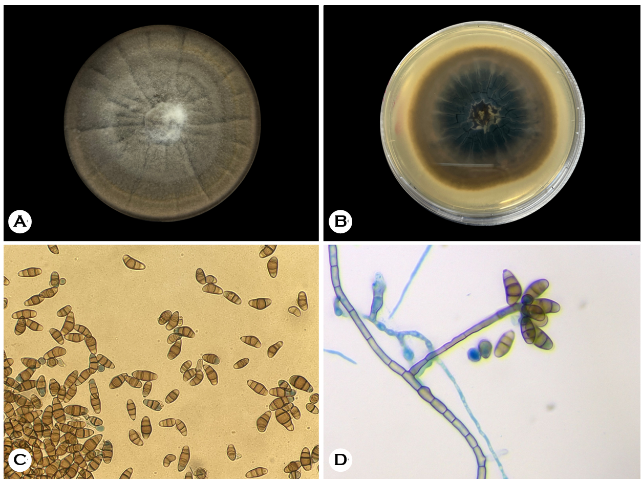
Curvularia clavata colonies have rapid growth; during the first 3 days, they show white colonies that later turn gray. On the fifth day, the colonies started turning olive green, and over time, they became black with a velvet-like texture and a fluffy center (Fig. 1A). The reverse side colonies are dark brown, with visible grooves and a clear halo at the periphery, without pigment production (Fig. 1B). Their optimal growth temperature is 20~25℃3. The conidia are smooth, either straight or curved, and have a claviform shape. Some cells are bigger than others and are colored pale to dark brown; apical and basal cells are both generally lighter. Their size ranges from 3~27 × 7~13.5 μm (Fig. 1C). The macrosiphon- ated mycelium with pigmented and segmented thick-wall hyphae are branched, pale to dark brown, with a diameter of 1.5~3.5 μm. Conidiophores can appear as isolated or in groups; commonly macronematous, they can also be observed as semi/mononematous. These conidiophores can be straight or curved, with cell walls thicker than vegetative hyphae, generally lighter at the top, and 34~102.5 × 2.5~5.5 μm2. Conidiogenous cells have smooth walls and can be in terminal or intercalary positions, proliferating sympodially. Their shape is subcylindrical or slightly swollen, being 5.5~17 × 4~6 μm2 (Fig. 1D).
For a more accurate description, molecular identification was performed by amplifying and sequencing the ribosomal internal transcribed spacer region. A sequence was deposited in GenBank under accession number PP373713.1.

References
1. van Vuuren N, Yilmaz N, Wingfield MJ, Visagie CM. Five novel Curvularia species (Pleosporaceae, Pleosporales) isolated from fairy circles in the Namib desert. Mycol Prog 2024;23:39
Google Scholar
2. Marin-Felix Y, Hernandez-Restrepo M, Crous PW. Multi-locus phylogeny of the genus Curvularia and description of ten new species. Mycol Prog 2020;19:559-588
Google Scholar
3. Madrid H, da Cunha KC, Gené J, Dijksterhuis J, Cano J, Sutton D, et al. Novel Curvularia species from clinical specimens. Persoonia 2014;33:48-60
Google Scholar
Congratulatory MessageClick here!