pISSN : 3058-423X eISSN: 3058-4302
Open Access, Peer-reviewed
pISSN : 3058-423X eISSN: 3058-4302
Open Access, Peer-reviewed

Jisoo Kim,Jisung Kim,Taekwoon Kim,Joonsoo Park
10.17966/JMI.2023.28.4.123 Epub 2024 January 05
Abstract
Keywords
Contaminants Fungus culture Penicillium spp
Fungal culture is a standard testing method for dermato- mycosis along with the KOH test. However, its positive rate is lower than that of the KOH test, and although it is very specific, it is less sensitive1,2. It is important to distinguish between contaminants and pathogens even after the fungus has been cultured.
Common fungal culture tests are performed on Sabouraud's dextrose agar and can be used with cycloheximide to prevent contamination3. However, a medium without cycloheximide must be used to cultivate fungi other than dermatophytes. The positive rate can be increased only when sufficient speci- mens are collected and cultured. During culturing, nonselec- tive and selective agar are simultaneously inoculated for samples from nonsterile locations or locations that may be contaminated with other microorganisms.
Scraping or curette inoculation on or into the agar at several points on the agar surface is the most suitable method for culture. The tissue must be mashed or minced and then uni- formly inoculated. If contamination does not occur and proper culture is performed, mold will grow at the inoculation site.
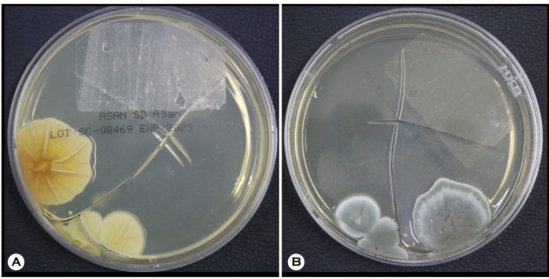
The photo below shows a case of contamination by Penicillium spp., which was confirmed using gross and micro- scopic findings. Cultures were conducted on three media plates, and the fungus was cultured only on the one shown in the photo (Fig. 1). It appears to be contaminated with an airborne fungus. When culturing fungi, it is important to take precautions to avoid confusing contaminants with pathogens.
References
1. Levitt JO, Levitt BH, Akhavan A, Yanofsky H. The sensitivity and specificity of potassium hydroxide smear and fungal culture relative to clinical assessment in the evaluation of tinea pedis: a pooled analysis. Dermatol Res Pract 2010;2010:764843
Google Scholar
2. Borman AM, Johnson EM. Interpretation of fungal cul- ture results. Curr Fungal Infect Rep 2014;8;312-321
Google Scholar
3. Iwern PC, Thomson GR, Wiederhold NP. Mycotic disease. In: McPherson RA, Pincus MR, editors. Henry's clinical diagnosis and management by laboratory methods. 24th ed. NewYork:Elsvier, 2022:1194-1228
Congratulatory MessageClick here!
