pISSN : 3058-423X eISSN: 3058-4302
Open Access, Peer-reviewed
pISSN : 3058-423X eISSN: 3058-4302
Open Access, Peer-reviewed

Jueon Roh,Jisoo Kim,Jisung Kim,Taekwoon Kim,Joonsoo Park
10.17966/JMI.2023.28.3.90 Epub 2023 October 06
Abstract
Keywords
Enterococcus durans Enterococcus hirae Green nail syndrome
Green nail syndrome is characterized by the green dis- coloration of the nail plate (ranging from greenish-yellow and greenish-brown to greenish-black), proximal chronic non- tender paronychia, and distolateral onycholysis. This condition is predominantly caused by a Pseudomonas aeruginosa infection in individuals whose nails are frequently exposed to water, soaps, detergents, or mechanical trauma1. The green discoloration observed in P. aeruginosa infections stems from the production of pyocyanin or fluorescein. However, various case reports have implicated numerous microorganisms, such as Klebsiella, Gram-positive bacteria, Aspergillus, and Candida spp., as causative organisms of green nail syndrome. We present a case of green nail syndrome caused by Gram-positive bacteria.
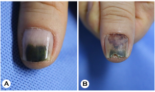
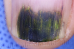
A 58-year-old male presented with navy green to greenish-brown nails on both thumbs that persisted for several months (Fig. 1A). Dermoscopy revealed a brown discoloration at the edge of the lesion (Fig. 2). The lesion initially appeared as a small coin-sized mark, which gradually enlarged and seemed to merge. He frequently exposed his hands to water and had a medical history of dyslipidemia and asthma. Other than those observed in both thumbnails, no specific dermatologic lesions were observed. Complete blood count and biochemistry findings were unremarkable, except for an elevated eosinophil count (6.9%). Biopsy samples were obtained from the left thumbnail, and gram staining and bacterial cultures were conducted on both thumbnails. Histo- pathology revealed focal hemorrhage, whereas D-PAS and gram staining showed no significant findings. However, nail scraping cultures from both thumbnails tested positive for Enterococcus durans and Enterococcus hirae. Considering the brownish discoloration on the edge of the nails, a diag- nosis of green nail syndrome induced by E. durans and E. hirae was established. Based on an antibiotic sensitivity test, treatment with oral minocycline (100 mg/daily) and mupirocin ointment was started. After 7 weeks, follow-up cultures confirmed bacterial absence, indicating improvement in the green nail syndrome (Fig. 1B).
Enterococci are Gram-positive, facultatively anaerobic organisms commonly found in the human intestinal flora. They typically cause urinary tract infections, bacteremia, endo- carditis, wound infections, and intra-abdominal infections. Enterococcal nail infections are rare, though some reports do exist2. These infections manifest as yellowish-brown dis- colorations likely due to flavonoids and carotenoids produced by the Enterococci3,4. In this particular case, we observed a navy green to greenish-brown central discoloration with brownish edges. This could have been attributed to the high density of pigment produced by the Enterococci. The confined space within the nail bed compared to other body parts might have caused the higher density of Enterococci. Hence, enterococcal nail infections may manifest as greenish dis- colorations, otherwise known as green nail syndrome.
In conclusion, P. aeruginosa should not be considered the sole causative agent for green nail syndrome. Performing not only bacterial culture but also KOH and fungal culture is necessary5. Patients should be treated appropriately according to culture results and antibiotic sensitivity tests.
References
1. Chiriac A, Brzezinski P, Foia L, Marincu I. Chloronychia: green nail syndrome caused by Pseudomonas aeruginosa in elderly persons. Clin Interv Aging 2015;10:265-267
Google Scholar
2. Ouzounova-Raykova VV. Green nail syndrome on the nail plate and bed related with Enterococcus and Fusarium coinfection. Folia Med 2022;64:547-550
Google Scholar
3. Maraccini PA, Ferguson DM, Boehm AB. Diurnal variation in Enterococcus species composition in polluted ocean water and a potential role for the Enterococcal carote- noid in protection against photoinactivation. Appl Environ Microbiol 2012;78:305-310
Google Scholar
4. Tayler RF, Ikawa M, Chesbro W. Carotenoids in yellow-pigmented Enterococci. J Bacteriol 1971;105:676-678
Google Scholar
5. Ohn J, Yu DA, Park H, Cho S, Mun JH. Green nail syn- drome: Analysis of the association with onychomycosis. J Am Acad Dermatol 2020;83:940-942
Google Scholar
Congratulatory MessageClick here!
