pISSN : 3058-423X eISSN: 3058-4302
Open Access, Peer-reviewed
pISSN : 3058-423X eISSN: 3058-4302
Open Access, Peer-reviewed

Sang-Kyung Lee,Geon-Jong Lee,Kyung-Hwa Nam,Jin Park
10.17966/JMI.2023.28.2.58 Epub 2023 July 07
Abstract
Keywords
Dermoscopy Microsporum canis Scanning electron microscopy Tinea capitis
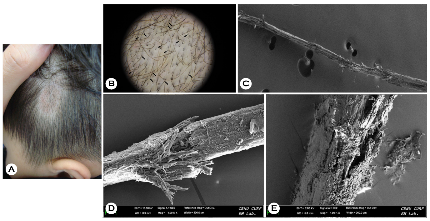
A 3-year-old boy presented with localized bald patches on his temporal scalp, which had been present for a month. He had been in close contact with a dog and his family and past medical histories were unremarkable. Physical examination revealed multiple round erythematous scaly patches with broken hair in the hairless area. Dermoscopic examination showed Morse code-like and sharp-angled zigzag hair with transverse white bands across the hair shaft (Fig. 1A). Scan- ning electron microscopy of the affected hair revealed thin and destroyed cuticles covered with numerous fungal arthro- conidia corresponding to the white bands on the hair shaft (Fig. 1B-1E). Microsporum canis was identified as the causative species via fungal culture and polymerase chain reaction. The patient received a treatment of oral griseofulvin (125 mg/day) and topical terbinafine for 8 weeks.
Dermatophyte infections of the hair can result in three distinct patterns of fungal invasion: ectothrix, endothrix, or favus, depending on the specific species involved1. Ectothrix tinea capitis, often associated with Microsporum canis, is characterized by the accumulation of arthroconidia around the exterior of the hair shaft, leading to the circumferential destruction of the hair cuticle2. As the infected hair continues to grow, the affected portion rises above the scalp surface and becomes more prone to bending or breaking due to increased fragility. This gives rise to the appearance of Morse code-like or bent zigzag hair with transverse white bands, indicating focal weakening of the hair shaft caused by fungal invasion3. Understanding this information can assist clinicians in predicting the type of fungal invasion and comprehending the formation mechanism behind the distinctive dermoscopic features observed in cases of ectothrix tinea capitis.
References
1. Elewski BE. Tinea capitis: a current perspective. J Am Acad Dermatol 2000;42:1-20
Google Scholar
2. Shelley WB, Shelley ED, Burmeister V. The infected hairs of tinea capitis due to Microsporum canis: demonstration of uniqueness of the hair cuticle by scanning electron microscopy. J Am Acad Dermatol 1987;16:354-361
Google Scholar
3. Meneses OM, Donati A, Silva FO, Mimiça MJ, Machado CJ, Veasey J. Trichoscopy patterns of tinea capitis and their correlation with mycological culture results. J Am Acad Dermatol 2023;88:166-167
Google Scholar
Congratulatory MessageClick here!
