pISSN : 3058-423X eISSN: 3058-4302
Open Access, Peer-reviewed
pISSN : 3058-423X eISSN: 3058-4302
Open Access, Peer-reviewed

Sang-Kyung Lee,Han-Nah Park,Yeon-Soo Kim,Jin Park
10.17966/JMI.2023.28.1.21 Epub 2023 April 05
Abstract
Keywords
Dermoscopy Scanning electron microscopy Tinea capitis Trichophyton tonsurans
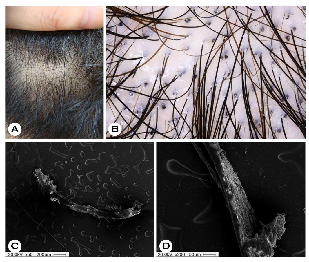
An 18-year-old female wrestler presented with a 1-month history of pruritic diffuse hairless patches on her occipital scalp. She had no contact history with animals, and her family and past medical histories were unremarkable. Physical examination revealed multiple short, broken hairs with minimal inflammation on the hairless area of the occiput (Fig. 1A). Dermoscopic findings indicated comma hairs, black dots, short, broken hairs, and mild scales (Fig. 1B). Scanning electron microscopy of the comma hairs revealed that the hair shaft was filled with numerous fungal spores (Fig. 1C-D). Fungal culture and PCR identified Trichophyton tonsurans as the causative species, so she was treated with 6 weeks of oral terbinafine (250 mg/day) and topical isoconazole.
Identifying the causative species of tinea capitis (TC) is important because the most appropriate treatment de- pended on the causal agent. Trichophyton species (usually with endothrix-type) and Microsporum species (usually with ectothrix-type) are the most common species that cause TC1. During endothrix Trichophyton infection, the fungus migrates through the hair follicles and penetrates the cortex. After the invasion of the fungus into the hair cortex, the transforming arthrospores occupy and weaken the inner root sheath, forming short, broken hairs at the scalp's surface, appearing as black dots. Dermoscopy provides an immediate diagnosis of TC and may predict the type of infection2. Trichophyton TC usually presents as comma and corkscrew hairs, while Microsporum TC exhibits morse-code-like hairs2,3. Microscopic examination of the infected hairs can confirm the diagnosis of TC and may establish the type of fungal invasion.
References
1. Elewski BE. Tinea capitis: a current perspective. J Am Acad Dermatol 2000;42:1-20
Google Scholar
2. Meneses OM, Donati A, Silva FO, Mimiça MJ, Machado CJ, Veasey J. Trichoscopy patterns of tinea capitis and their correlation with mycological culture results. J Am Acad Dermatol 2023;88:166-167
Google Scholar
3. Park J. Dermoscopy of superficial dermatomycosis. Korean J Med Mycol 2017;22:53-61
Google Scholar
Congratulatory MessageClick here!
