pISSN : 3058-423X eISSN: 3058-4302
Open Access, Peer-reviewed
pISSN : 3058-423X eISSN: 3058-4302
Open Access, Peer-reviewed

Yong Woo Choi,Nuri Na,Joonsoo Park
10.17966/JMI.2018.23.2.59 Epub 2018 July 01
Abstract
Keywords
The genus Cladosporium forms a large part of demati- aceous hyphomycetes1. The saprophyte Cladosporium sphaerospermum can be found in diverse environments such as indoor and outdoor air, soil, hypersaline water, paint, silicone, decaying vegetation, and textile1. C. sphaerospermum also frequently exists as an airborne contaminant1. This fungus causes subcutaneous phaeohyphomycosis, intrabronchial lesions, human corneal ulcers, and onychomycoses2. Most Cladosporium species do not grow at temperatures exceeding 35℃. In this analysis, we sampled the right eyebrow of a 51-year-old woman who was tattooed a month ago.
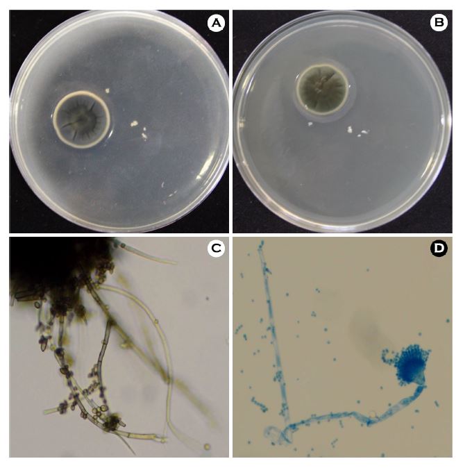
After streaking on Sabouraud dextrose agar, a flat, radially furrowed, crater-like colony was observed (Figure 1A). The colony was olivaceous green at the top and blackish green at the bottom (Figure 1B). Microscopically, the conidia of C. sphaerospermum were brown to dark brown in color and globose to subglobose in shape at their ends. The presence of ramoconidia is one of the characteristics of Cladosporium species3. The hyphae of C. sphaerospermum were septated with thick and darkened septa (Figure 1C). Lactophenol cotton blue staining of C. sphaerospermum revealed bluish branching septated hyphae with conidia chains (Figure 1D).
Conidia are formed via budding, with the youngest conidia at the tip and older conidia at the base. The youngest conidia are round in shape, whereas older conidia are elongated and septated. Because C. elatum, C. herbarum, and C. cladosporioides have similar appearances, sequence analysis is needed for their differentiation. When differentiating various species of dermatophytes, such as C. fusiforme, consideration should be given to the characteristic macroscopic and microscopic findings of C. sphaerospermum. Furthermore, because C. sphaerospermum is the most common contaminant, its presence should be confirmed by several cultures.
In relation to this article, I declare that there is no conflict of interest.
References
1. Bensch K. Braun U, Groenewald JZ, Crous PW. The genus Cladosporium. Stud Mycol 2012;72:1-401
Crossref
PubMed
2. Maduri A, Patnayak R, Verma A, Mudgeti N, Kalawat U, Asha T. Subcutaneous infection by Cladosporium sphaero- spermum-A rare case report. Indian J Pathol Microbiol 2015;58:406-407
Crossref
Google Scholar
PubMed
3. Schubert K, Groenewald JZ, Braun U, Dijksterhuis J, Starink M, Crous PW, et al. Biodiversity in the Cladosporium herbarum complex (Davidiellaceae, Capnodiales), with standardisation of methods for Cladosporium taxonomy and diagnostics. Stud Mycol 2007;58:105-156
Crossref
Google Scholar
Congratulatory MessageClick here!
