pISSN : 3058-423X eISSN: 3058-4302
Open Access, Peer-reviewed
pISSN : 3058-423X eISSN: 3058-4302
Open Access, Peer-reviewed

Hyun Ji Lee,Chihyeon Sohng,Jun Young Kim,Kyung Duck Park,Seok-Jong Lee,Weon Ju Lee
10.17966/JMI.2020.25.2.42 Epub 2020 July 04
Abstract
Keywords
Arthroconidia Chlamydospore Dermatophytosis Ex vivo human skin Histopathology Trichophyton rubrum
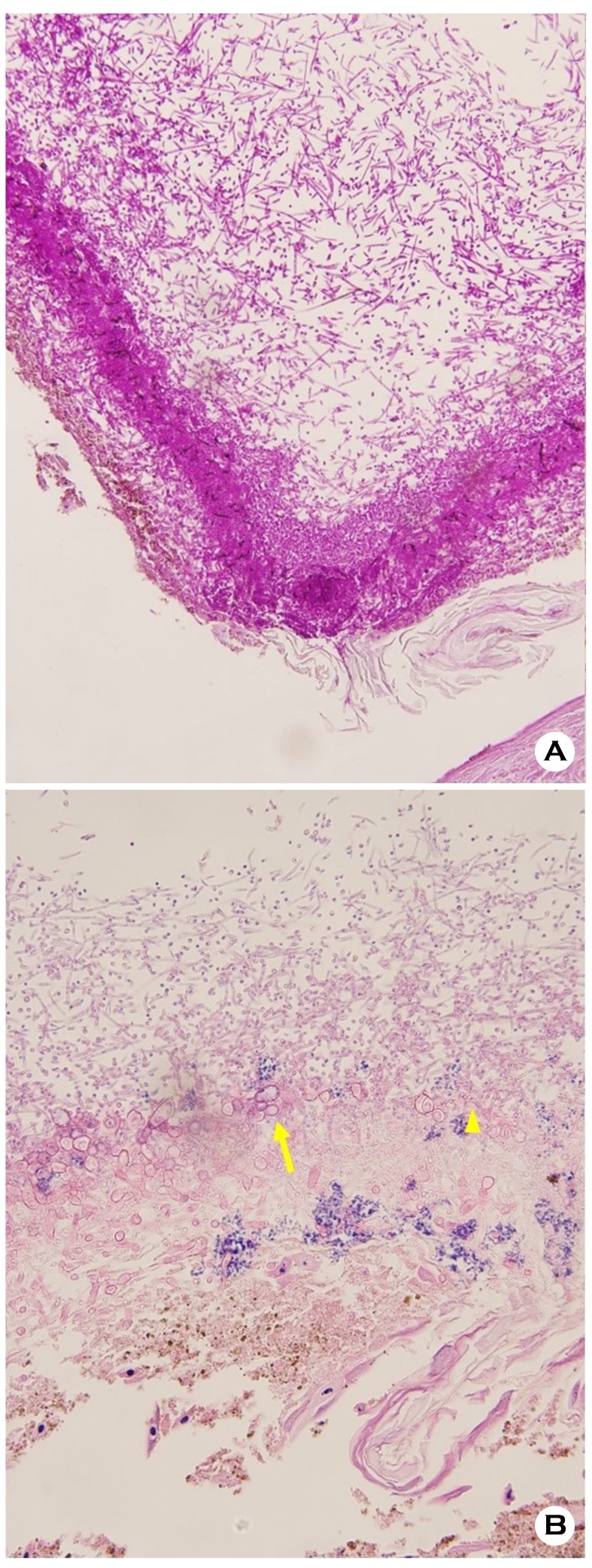
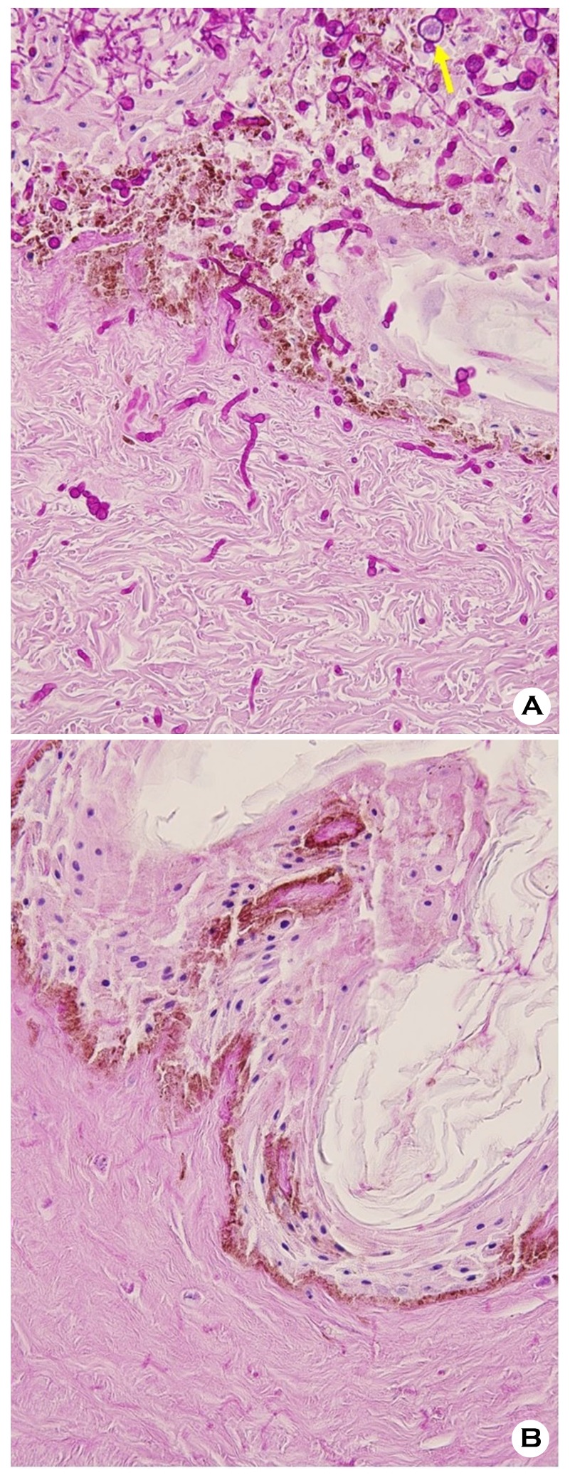
Trichophyton rubrum (T. rubrum) is the most usual dermatophyte that is responsible for dermatophytosis in humans1. A classic characteristic of dermatophytes is being keratinophilic. Thus, they occupy keratinized structures, including the stratum corneum of the skin, hairs, and nails. Occasionally, T. rubrum occupies the deeper skin or distant internal organ in immunosuppressed patients2,3. It is unfortunate that its pathogenic mechanism of invasion into the stratum corneum and dermis is not still totally understood. Different models, such as the animal model, stripped sheets of the stratum corneum, nail plates, monolayer cell culture model, or reconstructed human epidermis, were utilized for the examination of the mechanism of dermatophyte infection4-8. Even though they had a few limitations to imitate dermatophyte infections in humans, they can partly exhibit the pathogenic mechanism of dermatophyte infection. In our study, we investigated the histopathological characteristics in T. rubrum infection with the use of ex vivo human skin. Trichophyton rubrum was cultured for 2 weeks at 24℃ on Sabouraud dextrose agar. It was transferred to 10 ex vivo skin specimens which were foreskin acquired from circumcision. The ex vivo skin specimens were kept on the Sabouraud dextrose agar at 24℃ for 1 week. Afterward, histopathological examination was performed using the periodic acid-Schiff-diastase (PAS-D) stain under a light microscope. In the examination, slender septate hyphae with several arthroconidia were found in the stratum corneum (Figure 1A, 1B). Trichophyton rubrum often generates arthroconidia in vivo. Arthroconidia are involved in pathogenesis and function as a source of infection. Thicker septate hyphae and chlamydospore were found in the lower epidermis (Figure 1B, Figure 2A). Thicker septate hyphae and chlamydospore were also shown in the dermis (Figure 1B, Figure 2A, 2B). Chlamydospore is the life stage which survives in unfavorable conditions. Corzo-León et al.9 performed a study with ex vivo human skin placed on Dulbecco's Modified Eagle Medium. They exhibited long hyphae in the ex vivo skin surface 10 days following T. rubrum infection. Hyphae and arthroconidia of T. rubrum were much more demonstrated in ex vivo human skin in our study than in Corzo-León et al's. Liang et al.8 introduced the reconstructed human epidermis for T. rubrum infection model. In the said study, conidia and hyphae of T. rubrum were seen in the stratum corneum of the reconstructed human epidermis 4 days following its inoculation. Apart from this, infection with much more conidia of T. rubrum displayed a full epidermal invasion beyond the superficial keratinous layer. Ho et al.4 examined T. rubrum infection with the explanted porcine skin model. In the study, extended duration of infection in the skin led to luxurious growth and invasion of the dermis. The conditions might be the result of no active immune system that would restrain fungal growth, similar to our study. Invasive dermatophytoses happen more frequently in the immunocompromised host than in the healthy person. Trichophyton rubrum accounts for the majority of invasive fungal infections in immunosuppressed patients. Trichophyton rubrum in superficial dermatophytoses is confined in the stratum corneum, demonstrating a thin septate hyphae fragment in PAS staining. Nonetheless, it is possible for T. rubrum to show atypical and bizarre hyphae in the dermis in invasive dermatophytoses5. So, these findings make suitable diagnosis hard. Our study exhibited an atypical morphology of T. rubrum, such as chlamydospores and thicker hyphae, in the lower epidermis and dermis. These results will allow the recognition of the possibility of appearance changes of T. rubrum in invasive dermatophytoses.
References
1. Lee WJ, Kim SL, Jang YH, Lee SJ, Kim DW, Bang YJ, et al. Increasing prevalence of Trichophyton rubrum identified through an analysis of 115,846 cases over the last 37 years. J Korean Med Sci 2015;30:639-643
Google Scholar
2. Gong JQ, Liu XQ, Xu HB, Zeng XS, Chen W, Li XF. Deep dermatophytosis caused by Trichophyton rubrum: report of two cases. Mycoses 2007;50:102-108
Google Scholar
3. Squeo RF, Beer R, Silvers D, Weitzman I, Grossman M. Invasive Trichophyton rubrum resembling blastomycosis infection in the immunocompromised host. J Am Acad Dermatol 1998;39:379-380
Google Scholar
4. Ho FK, Delgado-Charro MB, Bolhuis A. Evaluation of an explanted porcine skin model to investigate infection with the dermatophyte Trichophyton rubrum. Mycopathologia 2020;185:233-243
Google Scholar
5. Aljabre SH, Richardson MD, Scott EM, Shankland GS. Germination of Trichophyton mentagrophytes on human stratum corneum in vitro. J Med Vet Mycol 1992;30:145 -152
Google Scholar
6. Yue X, Li Q, Wang H, Sun Y, Wang A, Zhang Q, et al. An ultrastructural study of Trichophyton rubrum induced onychomycosis. BMC Infect Dis 2015;15:532
Google Scholar
7. Firat YH, Simanski M, Rademacher F, Schröder L, Brasch J, Harder J. Infection of keratinocytes with Trichophytum rubrum induces epidermal growth factor-dependent RNase 7 and human beta-defensin-3 expression. PLoS One 2014;9:e93941
Google Scholar
8. Liang PP, Huang XZ, Yi JL, Chen ZR, Ma H, Ye CX, et al. A Trichophyton rubrum infection model based on the reconstructed human epidermis - Episkin®. Chin Med J (Engl) 2016;129:54-58
Google Scholar
9. Corzo-León DE, Munro CA, MacCallum DM. An ex vivo Human Skin Model to Study Superficial Fungal Infections. Front Microbiol 2019;10:1172
Google Scholar
Congratulatory MessageClick here!
