pISSN : 1226-4709 eISSN: 2465-8278
Open Access, Peer-reviewed
pISSN : 1226-4709 eISSN: 2465-8278
Open Access, Peer-reviewed

Yong Woo Choi,Jin Woo Park,Joonsoo Park
10.17966/JMI.2020.25.3.62 Epub 2020 October 06
Abstract
Keywords
Air crescent CT findings Fungal rhinosinusitis
Fungal rhinosinusitis (FRS) encompasses a wide variety of fungal infections, ranging from irritation to rapidly fatal in- fections1. Fungi colonize the nasal cavity and paranasal sinuses invasively or noninvasively2. The prevalence of FRS has been increasing over several decades2. There is evidence that the incidence of fungal ball is increasing because detection tech- niques and methods, such as radiologic evaluation, fungal culture techniques, and surgical technologies, have improved2. In FRS, computed tomography (CT) scans show opacity of the cavity associated with a hyperdense area within the lesion and thickened bone2. The most frequent fungus found in a fungal ball is Aspergillus fumigatus2. Removal of the mass with functional endoscopic sinus surgery is the treatment of choice for FRS2.
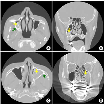
The typical CT findings of FRS are defined with spotted calcified plaque in the unilateral paranasal sinus, including the maxillary and sphenoid sinuses (Fig. 1 yellow arrow). Erosion of the sinus wall and enlargement of the sinus cavity also occurs. An axial CT view of a patient with FRS shows a filling defect in the maxillary sinus with thickened bone (Fig. 1. A, C). A coronal CT view of a patient with FRS shows sinus mucosal hypertrophy and hyperattenuation of calcified plaques (Fig. 1. B, D).
The incidence of FRS has been increasing recently due to environmental factors, condition of individual immune systems, and interaction of local tissue conditions3. It is difficult to distinguish between chronic rhinosinusitis and FRS by merely looking at the symptoms. Therefore, CT scan is a useful method for diagnosing FRS.
Patients provided written informed consent for the publication and the use of their images.
References
1. Chakrabarti A, Denning DW, Ferguson BJ, Ponikau J, Buzina W, Kita H, et al. Fungal rhinosinusitis: a categori- zation and definitional schema addressing current con- troversies. Laryngoscope 2009;119:1809-1818
Google Scholar
2. Liu X, Liu C, Wei H, He S, Dong S, Zhou B, et al. A retrospective analysis of 1,717 paranasal sinus fungus ball cases from 2008 to 2017. Laryngoscope 2020;130: 75-79
Google Scholar
3. Kim SW, Park YJ, Kim SW, Kang MG, Joo YH, Cho JH. A clinical analysis of fungal sinusitis. Korean J Otolaryngol 2005;48:332-337
Google Scholar
Congratulatory MessageClick here!
