pISSN : 3058-423X eISSN: 3058-4302
Open Access, Peer-reviewed

pISSN : 3058-423X eISSN: 3058-4302
Open Access, Peer-reviewed
Hyungrok Kim,Osung Kwon,Yong Joon Bang,Joonsoo Park
http://dx.doi.org/10.17966/KJMM.2016.21.3.103 Epub 2016 October 03
Abstract
Keywords
Trichophyton rubrum Fungal culture PDACT
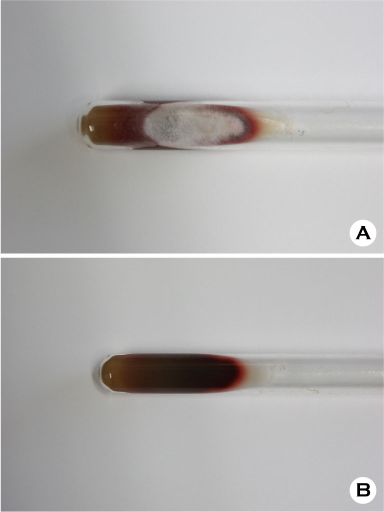
Trichophyton (T.) rubrum is an anthropophilic fungus also known as the most common dermatophyte of the human[1]. It is frequently associated with chronic skin infection involving nails and scalp. Morphologically, colonies present flat to slightly raised, white to reddish, downy to suede-like characteristics with either no reverse pigmentation or a yellow to wine-red reverse (Fig. 1A, 1B). Cultures usually show scanty slender clavate to pyriform microconidia. Macroconidia is typically absent. Prolonged cultured medium may show numerous chlamydospores with little clavate to pyriform microconidia.
The characteristic wine-red colored pigmentation on reverse is particularly intensified when the organism is grown on potato dextrose agar (PDA) or on corn meal agar with 1% dextrose. The corn meal and potato infusion provide essential nutrition base required for sporulation[2]. Dextrose serves as the energy source and is responsible for the reddish pigmentation. Utilization of tween 80 intensifies the reddish pigmentation which is conducive in differentiating the pathogen from Trichophyton mentagrophyte by KOH or saline assumption test[3]. In 1986, potato dextrose agar-corn meal-tween 80 (PDACT or "Chilgok plate") was developed at the Institute of Microbiology, Catholic skin clinic, Daegu, Korea, a suitable culture medium for T. rubrum. If culture fails to present reddish reverse pigmentation on media even in PDA or corn meal agar, subculture on PDACT may be a sound alternative in further evaluating the pathogen.

Dedications
This report is devoted to Dr. Soon-Bong Suh (1921-2007) Institute of Microbiology, Catholic skin clinic, Daegu, Korea, who strive to develop and generalize the use of the "Chilgok plate".
Conflict of interest
In relation to this article, I declare that there is no conflict of interest.
References
1. Suh SB, Kim KH, Bang YJ. Medical mycology. 1st ed. Seoul: Daihakseorim, 1994:45-48
2. Snyder JW, Atlas RM, LaRocco MT. Reagents, stains, and media: mycology. In: Versalovic J, Caroll KC, Jorgensen JH, et al. Manual of clinical microbiology. 10th ed. Washington DC: ASM Press, 2011:1767 -1775
3. Ates A, Ozcan K, Ilkit M. Diagnostic value of morphological, physiological and biochemical tests in distinguishing Trichophyton rubrum from Trichophyton mentagrophytes complex. Med Mycol 2008; 46:811-822
Google Scholar