pISSN : 3058-423X eISSN: 3058-4302
Open Access, Peer-reviewed
pISSN : 3058-423X eISSN: 3058-4302
Open Access, Peer-reviewed

Hyun-Min Seo,Ji Hun Park,Ki Yeon Kim,Joung Soo Kim
10.17966/JMI.2022.27.2.41 Epub 2022 July 01
Abstract
The patient provided written informed consent for the publication and the use of her images.
Keywords
Fungal melanonychia Terbinafine Ungual phaeohyphomycosis
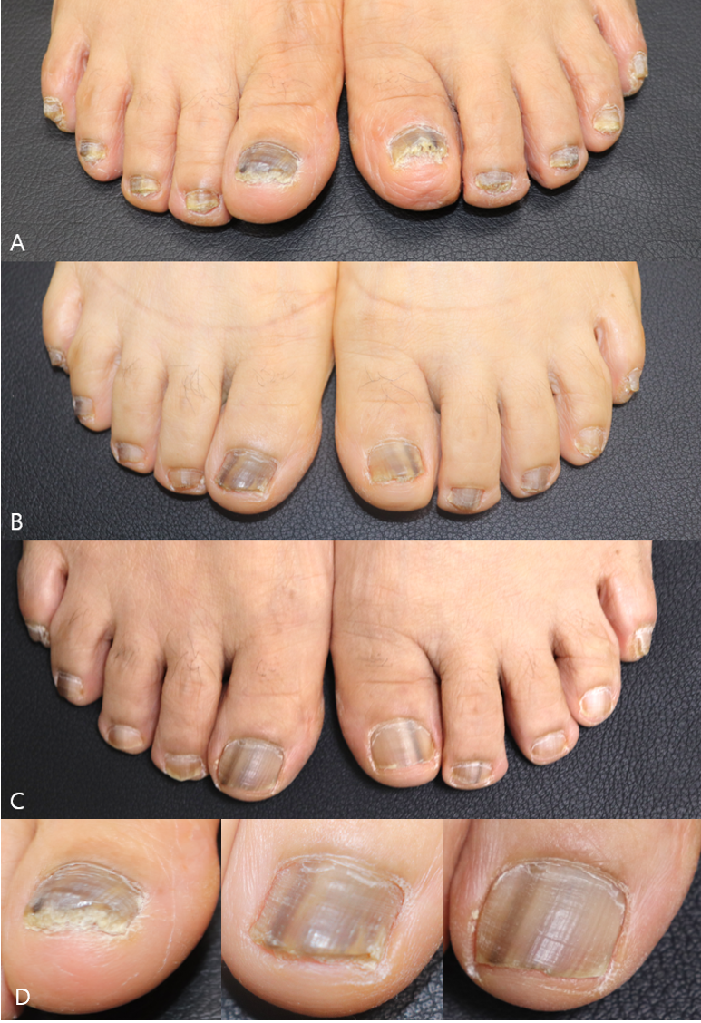
A 60-year-old female presented with multiple blackish pig- mentation on her toenails, along with nail change that first presented several months prior. Physical examination revealed multiple longitudinal blackish and yellowish striae on the first to fifth right and first and second left toenails. KOH test was performed on the involved toenails which revealed fungal hyphae. She was diagnosed with onychomycosis that induced melanonychia, known as fungal melanonychia (FM). The patient was treated with oral terbinafine for 9 months and she gradually improved (Fig. 1).
Fungal melanonychia (FM) or ungual phaeohyphomycosis is a rare nail disorder that presents with brownish or blackish pigmentation of the nail, mostly as a result of superficial in- fection of dematiaceous fungi1. There is a number of species that cause FM, the most common of which are Scytalidium dimidiatum and Trichophyton rubrum1. In a previous case series study, the clinical efficacy of oral terbinafine and itraconazole in patients with FM was reported2. However, there is no established treatment method for FM due to the lack of data and to the variability in responses to antifungal treatment. There are discussion regarding a species-specific causative approach. Our experience presents the clinical course of oral terbinafine treatment of FM with high-quality serial clinical photos. This can be helpful for the future management of patients with FM.
References
1. Finch J, Arenas R, Baran R. Fungal melanonychia. J Am Acad Dermatol 2012;66:830-841
Google Scholar
2. Lee SW, Kim YC, Kim DK, Yoon TY, Park HJ, Cinn YW. Fungal melanonychia. J Dermatol 2004;31:904-909
Google Scholar
Congratulatory MessageClick here!
