pISSN : 3058-423X eISSN: 3058-4302
Open Access, Peer-reviewed
pISSN : 3058-423X eISSN: 3058-4302
Open Access, Peer-reviewed

Dong Won Lee,Sangbin Jeong1,Moo Kyu Suh,Gyoung Yim Ha,Jong Im Lee
10.17966/JMI.2020.25.4.79 Epub 2021 January 06
Abstract
The patient provided written informed consent for the publication and the use of his images.
Keywords
Nocardia brasiliensis Nocardiosis Sporotrichoid
Nocardiosis is an uncommon infection caused by several species of bacteria belonging to the genus Nocardia (N), which are Gram-positive, partly acid-fast, filamentous, and branched bacilli. The infection may be classified into systemic and cutaneous types. Primary cutaneous nocardiosis is usually caused by N. brasiliensis, and may present in any one the following ways: 1) mycetoma, 2) lymphocutaneous (sporo- trichoid) infection, 3) localized superficial skin infection such as cellulitis, abscess, or granuloma, and 4) disseminated in- fection with cutaneous involvement. Sporotrichoid nocardiosis usually involves the upper extremities. Sulfamethoxazole is the drug of choice to treat this condition. There have been three reported cases of sporotrichoid primary cutaneous nocardiosis in Korean dermatological literature.
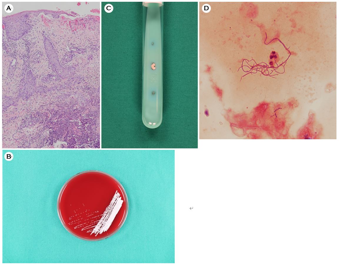
A 75-year-old male patient presented with painful skin lesions on his left arm of one-week duration. He recalled suffering a minor puncture injury to his hand while he was picking fruit. Examination of the skin revealed multiple erythematous ulcers and nodules on the left hand, forearm, and elbow (Fig. 1A). Abnormal laboratory test results included a total white blood cell (WBC) count of 9,460 cell/μL with 68% neutrophils. No other abnormal findings were revealed on the chest X-ray, electrocardiograph, blood biochemistry, or urine examination. Biopsy of the skin lesions and histopath- ological examination showed mild perivascular and interstitial infiltrate of lymphocytes and neutrophils (Fig. 2A). Culture of the biopsy material on blood agar and Ogawa medium at 37℃ was performed. The cultured organisms appeared as whitish, wrinkled, dry colonies on the blood agar plate after two days of incubation (Fig. 2B), and whitish, wrinkled, heaped up, dry colonies on 3% Ogawa medium at 37℃ after five days of incubation (Fig. 2C). Fungal culture was negative. Gram stain of the cultured colonies showed Gram-positive branching bacilli (Fig. 2D). The 16S ribosomal RNA gene sequencing of the clinical isolate was 99% identical to that of N. brasiliensis (GenBank accession number NR 041860.1). Following these investigations, the patient was diagnosed with primary sporotrichoid cutaneous nocardiosis caused by N. brasiliensis. He received trimethoprim/sulfamethoxazole 160/800 mg twice daily for two months as definitive treatment of his Nocardia infection. At his two-month follow up, he was noted to have made a complete recovery (Fig. 1B).
Sporotrichoid infection of nocardiosis can be misdiagnosed as sporotrichosis, cellulitis, and mycobacterial infection. We report the case of a patient with primary sporotrichoid cutaneous nocardiosis which was different from the usual clinical presentation of cutaneous nocardiosis. We have reported this case in order to emphasize and highlight the importance of histological examination, culture (bacterial, fungal, and myco-bacterial), and molecular analysis to enable correct diagnosis and treatment in atypical and difficult cases.

References
1. Lee SH, Suh CW, Choi JH, Sung KJ, Moon KC, Koh JK. A case of primary cutaneous sporotrichoid nocardiosis caused by Nocardia asteroides. Ann Dermatol 1999;11: 90-93
Google Scholar
2. In SG, Han SH, Shin JH, Choi GS, Chung MH. A case of disseminated nocardiosis secondary to the skin nodules in an elderly woman. Ann Dermatol 2008;20:82-85
3. Ryu HW, Lee KS, Rhyoo NH, Cho JW. Primary cutaneous nocardiosis with sporotrichoid pattern by Nocardia bra- siliensis in lung cancer patient. Korean J Dermatol 2012; 50:468-471
Google Scholar
4. Kang GS, Kim DM, Lee MH, Suh MK, Ha GY, Jang TJ, et al. Primary cutaneous nocardiosis caused by Nocardia brasiliensis. Korean J Dermatol 2011;49:730-734
Google Scholar
5. Shin JU, Kwon YS, Kim HJ, Park YJ, Lee KW, Lee KW. A case of disseminated cutaneous nocardiosis due to Nocardia brasiliensis diagnosed by fine needle aspiration biopsy and 16S ribosomal RNA sequencing. Korean J Dermatol 2009;47:1024-1029
Google Scholar
Congratulatory MessageClick here!
