pISSN : 3058-423X eISSN: 3058-4302
Open Access, Peer-reviewed
pISSN : 3058-423X eISSN: 3058-4302
Open Access, Peer-reviewed
Sang Youl Yun,Min Woo Park,Moo Kyu Suh,Gyoung Yim Ha
http://dx.doi.org/10.17966/KJMM.2016.21.4.129 Epub 2016 December 27
Abstract
Onychomycosis is caused by dermatophytes usually, but some species of nondermatophytic molds and yeasts are also associated with invasion of nails. Aspergillus(A.) terreus is a nondermatophytic mold which is opportunistic filamentous fungus in all environments. We report a case of onychomycosis caused by A. terreus in a 60-year-old male. The patient showed brownish yellow discoloration with hyperkeratotic change on the distal and lateral portion of both toenails. Direct microscopic examination of scraping on the potassium hydroxide preparation revealed septate hyphae and repeated cultures on Sabouraud's dextrose agar showed the velvety, cinnamon brown colonies. Biseriate and compactly columnar phialides that cover upper vesicle with conidial structure were shown in the slide culture. The DNA sequence of internal transcribed spacer (ITS) region of clinical sample was 99% match to that of A. terreus strain ATCC
20542 (GenBank accession number GU256759.1). We confirmed A. terreus by KOH mount, colony, light microscopic morphology and DNA sequence analysis. The patient was treated with 200 mg oral itraconazole daily and topical 5% amorolfine nail lacquer for 3 months.
Keywords
Aspergillus terreus Toenail onychomycosis
서 론
손발톱진균증은 손발톱의 진균성 감염증으로 그 원인균은 피부사상균(dermatophytes)이 대부분 이지만 비피부사상균성 사상균(nondermatophytic mold)이나 효모균(yeasts)에 의해 유발되기도 한다. 손발톱진균증을 야기하는 비피부사상균성 사상균으로 Aspergillus 균종, Scopulariopsis 균종, Fusarium균종, Acremonium 균종 등이 있다[1]. 이러한 균들은 오염균이나 기회감염균으로 간주되어 쉽게 간과되어 왔지만 최근의 면역기능의 저하를 가져오는 많은 질환 및 환경적 변화와 더불어 중요성이 증가되고 있다. 최근 비피부사상균성 사상균에 의한 손발톱진균증의 유병율은 점점 증가되고 있으며 이것은 이런 균들을 잠재적으로 원인균으로 보는 인식의 증가와 진단적 기술의 발달과 관련이 있다고 알려져 있다[2]. 손발톱진균증 중 Aspergillus균종에 의한 손발톱진균증은 국외에서 보고자에 따라 2.6~6.1%로 다양하게 보고되고 있다[3],[4],[5],[6]. 하지만 국내 피부과 문헌상 A. terreus에 의한 손발톱진균증은 아직 보고된 바가 없다.
이에 저자들은 60세 남자 발톱진균증 환자에서 임상, 진균배양, 광학 현미경 소견 및 분자생물학적 분석으로 A. terreus에 의한 발톱진균증으로 진단하고 드문 증례로 생각되어 문헌고찰과 함께 보고한다.
증 례
환 자: 김 O O, 60세, 남자
주 소: 양측 발의 소양감을 동반한 인설성 홍반성 반과 우측 첫 번째, 네 번째, 다섯 번째, 좌측 첫 번째, 두 번째, 네 번째, 다섯 번째 발톱의 황갈색 변색
현병력: 약 10년 전부터 발톱의 황갈색 변색이 있었고, 양측 첫 번째 발톱의 원위외측부에서는 비후된 소견을 보였음
과거력: 3년 전부터 지속되는 당뇨로 치료 중임
가족력: 특이 사항 없음
이학적 소견: 피부 소견 외 특이 사항 없음
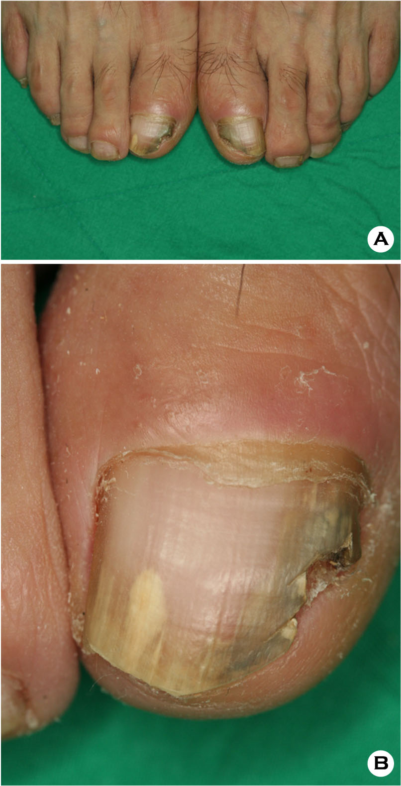
피부 소견: 양측 발에 인설성 홍반성 반과 우측 첫 번째, 네 번째, 다섯 번째, 좌측 첫 번째, 두 번째, 네 번째, 다섯 번째 발톱의 황갈색 변색이 보이고 양측 첫 번째 발톱의 원위부는 비후된 소견을 보였음(Fig. 1A, 1B).
검사실 소견: 일반혈액 및 말초혈액도말검사, 대소변검사, 매독혈청검사, 간기능 및 신기능 검사, 간염 항원 항체 검사, HIV 검사, 흉부 X-선 검사, 심전도는 모두 정상범위 내지 음성을 보였다.
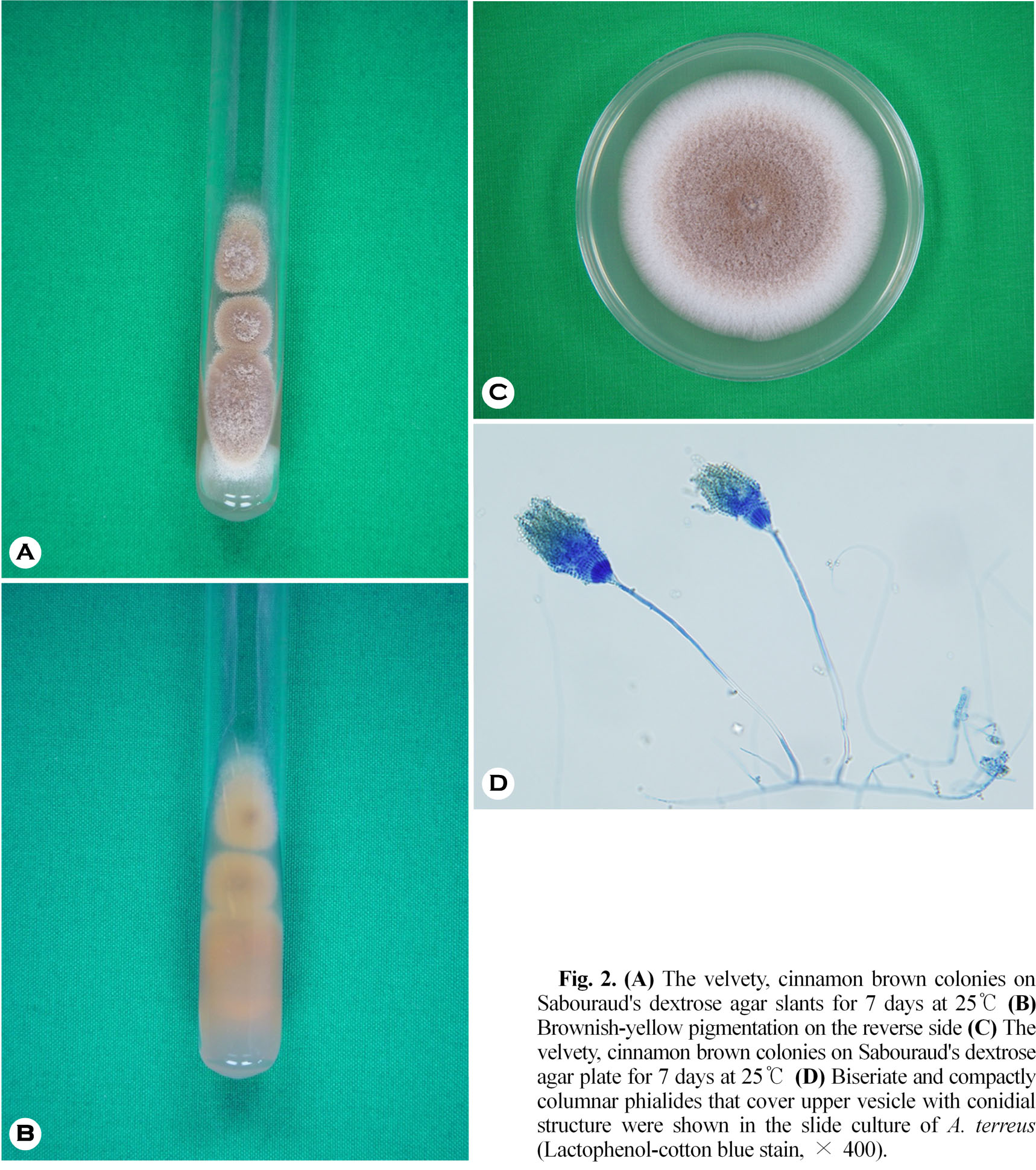
진균학적 소견: 병변부 손발톱의 KOH 검사상 분절균사를 관찰할 수 있었으며, 사부로 사면배지에 접종하여 25℃에서 1주간 배양한 결과 3개의 동일한 벨벳 같은 계피색의 균집락이 관찰되었고,배지의 뒷면은 황갈색의 착색이 보였고, 평판배지에 계대배양 하였을 때도 동일한 소견을 보였다(Fig. 2A, 2B, 2C). 이 집락을 슬라이드 배양표본을 만들어 lactophenol cotton blue로 염색하여 현미경 관찰상 유리질의 격막균사(hyaline septate hyphae)를 보이며, 구상의 정낭(vesicle)과 치밀한 원주상의 2단 경자(biseriated phialides)의 끝에 분생포자가 구상의 상부을 덮고 있으며, 분생포자를 관찰할 수 있었다(Fig. 2D).
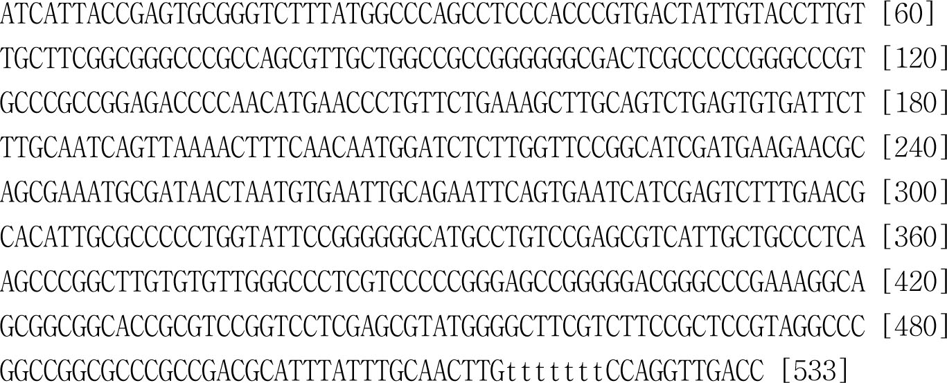
분자생물학적 분석: 환자의 배양된 균집락으로 부터 DNA를 분리하여 진균핵 내의 internal transcribed spacer (ITS) 부위의 염기서열을 얻은 후 Blast를 이용하여 GenBank에 있는 A. terreus strain ATCC 20542 (GenBank accession number GU-256759.1)의 ITS 부위의 염기서열과 비교한 결과 99% 일치하였다(Fig. 3). 이상의 KOH 소견, 진균 배양, 광학 현미경 소견, 그리고 ITS 부위의 염기서열 분석으로 A. terreus로 동정하였다.
치료 및 경과: 3개월간 itraconazole 1일 200 mg경구 투여와 5% amorolfine nail lacquer의 도포로 치료하였다.
고 찰
손발톱진균증은 피부사상균, 비피부사상균성 사상균, 그리고 효모균 등에 의해 유발되는 손발톱의 진균성 감염증으로 전체 손발톱질환의 약20%를 차지하는 흔한 감염증이다[1]. 피부사상균을 제외한 비피부사상균성 사상균에 의한 손발톱진균증은 손발톱진균증의 1.45~17.6%를 차지하며, Aspergillus 균종, Scopulariopsis 균종, Fusarium 균종, Acremonium 균종 등이 원인이 될 수 있다[5],[7],[8],[9],[10],[11].
Aspergillus 균종에 의한 손발톱진균증은 아마도 Aspergillus 균종이 피부사상균과는 달리 각질친화적(keratonophilic)이지 않기 때문에 족부 외상이나 또는 다양한 이전의 선행질환 후 이차적인 감염에 의해서 초래되지만[12], 몇몇 연구들은 손발톱진균증의 1차 원인균으로 Aspergillus 균종을 보고하였다[13],[14]. Aspergillus 균종에 의한 손발톱진균증은 국외에서 보고자에 따라 2.6~6.1%로 다양하게 보고되고 있으며[3],[4],[5],[6], 국내에서는 Aspergillus 균종에의한 손발톱진균증은 Lim 등7에 의해서 4.6%로 보고되고 있다. 이처럼 많은 차이를 보이는 이유는 비피부사상균성 사상균의 분포의 지역적 차이, 비피부사상균성 사상균에 의한 손발톱진균증의 진단에 있어서 다른 기준의 적용, 비피부사상균성사상균의 배양에 있어서 부적절한 진균학적 방법의 사용 등에 기인한다고 한다[8].
A. terreus는 유리질의 진균으로 전 세계적으로 토양과 공기 중에서 발견되며, 기회감염균인 A. terreus는 전신 및 표재성 감염을 일으킬 수 있고,손발톱진균증 중 가장 흔하게 첫 번째 발가락을 침범한다[15]. Hwang 등[11]은 A. terreus에 의한 손발톱진균증은 비피부사상균성 사상균에 의한 손발톱진균증의 5.1%를 차지하는 것으로 보고하였다.
Aspergillus 균종은 오염이 가능한 균이므로 감염된 손발톱에서 시행한 배양에서 Aspergillus 균종이 동정되었을 때 이것이 원인균인지 또는 단지 오염균인지 구별하는 것은 어렵다. 그래서 1976년 English[16]는 비피부사상균성 사상균이 배양되면 현미경 검사에서 균사, 또는 포자가 발견되고, 비피부사상균성 사상균에 의한 감염은 반복적인 배양검사에서 같은 균주가 동정될 때만 손발톱진균증의 원인균으로 간주할 수 있다고 하였으며, 최근에는 균주의 진균핵내 ITS 부위의 염기서열 분석이 진단이 어려운 손발톱진균증의 확진에 보조적으로 유용하게 사용되고 있다[12].
비피부사상균성 사상균인 A. terreus는 Aspergillus 균종에서와 같이 KOH 검사상 분절균사를 관찰할 수 있다[15],[17]. 이 균집락을 lactophenol cotton blue 염색하여 현미경으로 관찰하면 유리질의 격막균사를 보이고, 구상의 정낭(vesicle)과 치밀한 원주상의 2단 경자의 끝에 분생포자가 구상의 상부을 덮고 있다[15],[17],[18]. 본 증례에서도 KOH 검사에서 분절균사가 보였고, 반복적인 배양검사에서 동일한 배양 소견을 보였으며, lactophenol cotton blue 염색에서 동일한 현미경 소견이 관찰되었다. 또한 균집락으로부터 DNA를 추출하여 진균핵 내의 ITS 부위의 염기서열을 GenBank에 있는 A. terreus strain ATCC 20542 (GenBank accession number GU256759.1)의 ITS 부위의 염기서열과 비교한 결과 99% 일치하여 원인균주로 A. terreus로 동정하였다.
손발톱진균증의 임상형을 보면 1998년 Baran등[19]은 손발톱진균증을 원위외측 손발톱하 손발톱진균증(distal and lateral subungual onychomycosis,DLSO), 표재성 백색 손발톱진균증(superficial white onychomycosis, SWO), 근위 손발톱하 손발톱진균증(proximal subungual onychomycosis, PSO), 손발톱내 손발톱진균증(endonyx onychomycosis, EO), 전이상성 손발톱진균증(total dystrophic onychomycosis, TDO)으로 5가지 임상형으로 분류하였다. 이 중 손발톱진균증의 가장 흔한 유형은 DLSO이고[1],[7], 본 증례도 임상형이 DLSO였다. 비피부사상균성사상균에 의한 손발톱진균증은 일반적으로 치료에 잘 반응하지 않는 것으로 알려져 있으며[20], Tosti 등[8]은 Aspergillus 균종에 의한 손발톱진균증 7예 중 치료받은 5예서 경구 및 국소 치료로 모두 완치를 관찰하여 다른 비피부사상균성 사상균과는 달리 Aspergillus 균종에 의한 손발톱진균증은 치료에 잘 반응한다고 하였으며, 본 증례에서도 3개월간 itraconazole 1일 200 mg 경구투여와 함께 5%amorolfine nail lacquer 국소 도포를 병용함으로서 임상 및 진균학적 완치를 관찰할 수 있었다.
이에 저자들은 족부백선이나 수부백선의 동반이 없이 첫 번째 발톱의 원위외측부에 황갈색의 비후가 보이고 KOH 검사에서 균사가 관찰되며 항진균제에 비교적 잘 반응하는 비피부사상균성 손발톱진균증의 경우 A. terreus를 원인균으로 의심해 보아야 할 것으로 생각한다.
Conflict of interest
In relation to this article, I declare that there is no conflict of interest.
References
1. Schieke SM, Garg A. Superficial fungal infection. In Goldsmith LA, Katz SI, Gilchrest BA, Paller AS, Leffell DJ, Wolff K, editors. Fitzpatrick's dermatology in general medicine. 8th ed. New York: McGraw-Hill, 2012:2278-2297
2. Gupta AK, Ryder JE, Baran R, Summerbell RC. Nondermatophyte onychomycosis. Dermatol Clin 2003; 21:257-268
3. Romano C, Gianni C, Difonzo EM. Retrospective study of onychomycosis in Italy: 1985-2000. Mycoses 2005;48:42-44
Crossref
Google Scholar
4. Gupta M, Sharma NL, Kanga AK, Mahajan VK, Tegta GR. Onychomycosis: Clinico-mycologic study of 130 patients from Himachal Pradesh, India. Indian J Dermatol Venereol Leprol 2007;73:389-392
Crossref
Google Scholar
5. Gianni C, Romano C. Clinical and histological aspects of toenail onychomyosis caused by Aspergillus spp.: 34 cases treated with weekly intermittent terbinafine. Dermatology 2004;209:104-110
Crossref
Google Scholar
6. Hilmioqlu-Polat S, Metin DY, Inci R, Dereli T, Kilinc I, Tumbay E. Non-dermatophytic molds as agents of onychomycosis in Izmir, Turkey - a prospective study. Mycopathologia 2005;160:125-128
Crossref
Google Scholar
7. Lim SW, Suh MK, Ha GY. Clinical features and identification of etiologic agents in onychomycosis. Korean J Dermatol 2004;42:53-60
Google Scholar
8. Tosti A, Piraccini BM, Lorenzi S. Onychomycosis caused by nondermatophytic molds: clinical features and response to treatment 59 cases. J Am Acad Dermatol 2000;42:217-224
Crossref
Google Scholar
9. Gianni C, Cerri A, Crosti C. Non-dermatophytic onychomycosis. An underestimated entity. A study of 51 cases. Mycoses 2000;43:29-33
Crossref
Google Scholar
10. Summerbell RC, Kane J, Krajden S. Onychomycosis, tinea pedis and tinea manuum caused by nondermatophytic filamentous fungi. Mycoses 1989;32: 609-619
Crossref
Google Scholar
11. Hwang SM, Suh MK, Ha GY. Onychomycosis due to nondermatophytic molds. Ann Dermatol 2012;24:175 -180
Crossref
Google Scholar
12. Takahata Y, Hiruma M, Sugita T, Muto M. A case of onychomycosis due to Aspergillus sydowii diagnosed using DNA sequence analysis. Mycoses 2008;51:170 -173
Crossref
Google Scholar
13. Tosti A, Piraccini BM. Proximal subungual onychomycosis due to Aspergillus niger: report of two cases. Br J Dermatol 1998;139:156-157
Google Scholar
14. Grover S. Clinicomycological evaluation of onychomycosis at Banglore and Jorhat. Indian J Dermatol Venerol Leprol 2003;69:284-286
Crossref
Google Scholar
15. Fernandez MS, Rojas FD, Cattana ME, Sosa MDE L, Mangiaterra ML, Giusiano GE. Aspergillus terreus complex: an emergent opportunistic agent of onychomycosis. Mycoses 2013;56:477-481
Crossref
Google Scholar
16. English MP. Nails and fungi. Br J Dermatol 1976;94: 697-701
Crossref
Google Scholar
17. Kwon-Chung KJ, Bennett JE. Medical mycology. Philadelphia: Lea & Febiger, 1992:201-247
18. De Hoog GS, Guarro J, Gene J, Figueras MJ. Atlas of clinical fungi. 2nd ed. Virgili, Centraalbureau voor Schimmelcultures, 2000:442-519
19. Baran R, Hay RJ, Tosti A, Haneke E. A new classification of onychomycosis. Br J Dermatol 1998;139: 567-571
Crossref
Google Scholar
20. Nolting S, Brautigam M, Weidinger G. Terbinafine in onychomycosis with involvement by nondermatophytic fungi. Br J Dermatol 1994;130(Suppl 43):16 -21
Crossref
Google Scholar
