pISSN : 3058-423X eISSN: 3058-4302
Open Access, Peer-reviewed
pISSN : 3058-423X eISSN: 3058-4302
Open Access, Peer-reviewed

Jun Suk Hong,Dong Won Lee,Moo Kyu Suh,Gyoung Yim Ha,Jong Im Lee,Soyoun Shin
10.17966/JMI.2018.23.3.79 Epub 2018 September 28
Abstract
Keywords
Cutaneous infection Mycobacterium chelonae
Mycobacterium chelonae is an atypical rapidly growing Runyan Class IV Mycobacterium. It is widely distributed in soil and water, and causes skin infections after skin trauma or surgery1-3. Atypical mycobacteriosis can be difficult to diagnose because bacilli may not be obvious in tissue biopsies. Because pathogens differ in antimicrobial susceptibility, clinical suspicion of atypical mycobacterial infection should be confirmed through antimicrobial susceptibility testing and bacterial cultures2,4. Nine cases of M. chelonae infection have been reported in Korean dermatological literature previously, including cases of subcutaneous abscess after epidermal cyst incision and drainage, infection after injection of wrinkle fillers, acupuncture site infection, and infection after autologous fat transplantation. Only two of these cases did not have a trauma history5.
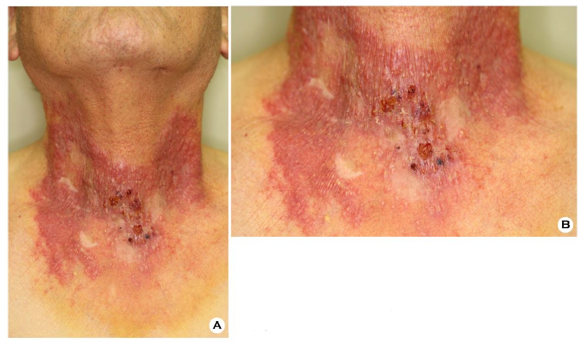
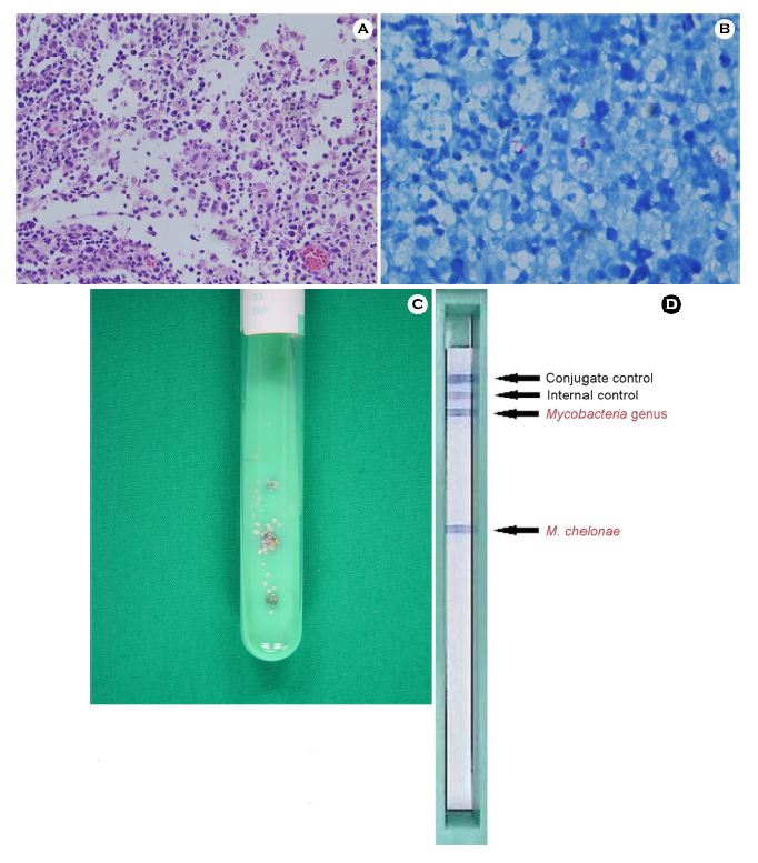
A 79-year-old man presented with multiple discrete or confluent pruritic, erythematous plaques with ulceration on the anterior neck that were of 3-weeks duration (Figure 1A, Figure 1B). He had a history of medication use for diabetes mellitus and no history of trauma-related skin lesions. There were no abnormal findings other than the cutaneous lesions. Histological evaluation indicated chronic inflammation with ill-defined granulomas and multinucleate giant cells in the deep dermis (Figure 2A). Filamentous bacilli were observed on acid-fast staining (Figure 2B), and 7-day cultures of skin biopsy specimens on 3% Ogawa medium yielded whitish, nonchromogenic, moist colonies (Figure 2C). The microorganism was identified as M. chelonae by using the AdvanSureTM Mycobacteria GenoBlot Assay kit (LG Life Sciences, Seoul, Korea (Figure 2D). Antimicrobial susceptibility testing showed sensitivity to amikacin, clarithromycin, and trimethoprim/sulfamethoxazole. The skin lesions completely resolved following the administration of 500 mg oral clarithromycin twice a day for 6 months. The cause of skin lesions in this 79-year-old man was diagnosed to be an M. chelonae infection based on clinical and pathological findings, positive skin culture, and genetic analysis. This rare case of M. chelonae infection is significant because of the absence of a history of skin trauma.
In relation to this article, I declare that there is no conflict of interest.
References
1. Kenneth ES, Oxman MN. Tuberculosis and infections with atypical mycobacteria, In: Wolff K, Goldsmith LA, Katz SI, Gilchrest BA, Paller AS, Leffel DJ, Dallas NA, editors. Fitzpatrick's dermatology in general medicine. 8th ed. New York: McGraw-Hill, 2012;2225-2241
Crossref
2. Han XY, De I, Jacobson KL. Rapidly growing mycobacteria: clinical and microbiologic studies of 115 cases. Am J Clin Pathol 2007;128:612-621
Crossref
Google Scholar
3. Gonzales-Santiago TM, Drage LA. Nontuberculous mycobacteria: skin and soft tissue infections. Dermatol Clin 2015;33:563-577
Crossref
Google Scholar
4. Chung WK, Park GH, Chang SE, Lee MW, Choi JH, Moon KC, et al. A case of Mycobacterium chelonae infection with foreign body granuloma after injection of filter. Korean J Dermatol 2008;46:1521-1525
Crossref
Google Scholar
5. Ahn HH, Park SD, Kim KM, Park CJ, Kim HU, Kim JP, et al. Infectious skin diseases, In: Textbook of dermatology. 6th ed. Seoul: Medbook, 2014;409-412
Crossref
Congratulatory MessageClick here!
