pISSN : 3058-423X eISSN: 3058-4302
Open Access, Peer-reviewed
pISSN : 3058-423X eISSN: 3058-4302
Open Access, Peer-reviewed

Hyungrok Kim,Dong Rak Kwon,Joonsoo Park
10.17966/JMI.2018.23.3.68 Epub 2018 September 28
Abstract
Background: Electric stimulation has been investigated for potential medical uses. Numerous articles have been published that focused on antimicrobial effects of electric current, but few studies have reported regarding modifications of fungal growth following exposure to electric current.
Objective: To evaluate effects of low alternating current on the growth of Trichophyton rubrum.
Methods: In total, 35 plates inoculated with T. rubrum were allocated to one of the five treatment groups (groups A, B, C, D, or E). Fungal colonies in each group were treated with a different intensity of electric current (0.5 μA, 4 μA, 25 μA, 600 μA, or 900 μA) at a frequency of 8 Hz. The area of each fungal colony was measured every other day for 7 days to evaluate the effects on fungal growth.
Results: No experimental groups treated with electric current showed any statistically significant differences against the control groups.
Conclusion: Microcurrent did not show any detectable changes in the viability of the fungus. Our findings indicate that microcurrent may affect fungal seeding to the media rather than the growth rate. Unfortunately, there are limited studies on this topic, and further research is warranted to clarify the precise effect of electric stimulation on the activity of microorganisms.
Keywords
Antifungal Antimicrobial Electric stimulus Fungal growth Microcurrent Trichophyton rubrum
The majority of superficial fungal infections are caused by dermatophytes, which belong to one of the following three genera: Epidermophyton, Trichophyton, and Microsporum1. Among these filamentous keratinophilic fungi, T. rubrum has been characterized as the most common pathogen of tinea unguium, tinea corporis, tinea pedis, and tinea capitis2. Treatment failures or drug resistance are common despite the administration of appropriately selected medications. Moreover, treating dermatophytes can be hindered by various factors, including the nonspecific spectrum of drugs activities, lack of efficacy, multiple drug interactions, and inadequate duration of medical treatment, resulting in frequent recurrences of the infection3. Different therapeutic strategies have been considered to improve chronic conditions of dermatophytosis. A few treatment modalities administered followed by the use of photodynamic devices, laser ablation, and ultra-violet irradiation have been reported to show some success in tackling chronic dermatophytosis4-6.
Delivery of weak electric currents through conductive electrodes has achieved inhibitory effects on microbial growth7-9 although there have been few studies on the effect of electrical stimulation on members of the Trichophyton genus. Recently, some studies have posited modifications of fungal growth through electrical current stimulation. An in vitro study by Kwon et al.10 showed faster growth of T. rubrum when 0.5, 2, or 4 μA of alternating current was applied via stainless steel electrodes for 30 min. In contrast, an in vitro experimental study by Novickij et al.11 demonstrated a marked irreversible growth inhibition of Candida albicans in response to exposure to electrical stimulation. The aim of our study was to evaluate the effect of low alternating current on the growth of T. rubrum.
1. Materials
1) Fungal strain, spore suspension preparation, and culture conditions
T. rubrum (Castellani) Sabouraud (ATCC 28188) was used in the study. The culture medium was obtained from the Catholic Skin Clinic, Daegu, Republic of Korea. Nutrient agar comprised potato dextrose agar, corn meal, and Tween 80 (PDACT) with added peptone. To prevent bacterial contamination, antibiotics were added to the medium (chloram-phenicol 500 mg L-1 and cycloheximide 500 mg L-1). 100 μl of inoculums derived from the spore suspension were subcultured on PDACT plates in 1.5 x 1.5 cm diameter and incubated for 3 days. A spore suspension was prepared by adding 10 mL sterile distilled water into a 1-week-old culture of T. rubrum, and the spore suspension was collected by gently withdrawing the liquid using a sterile pipette. The cell density of suspension was estimated to be 2.2 x 10⁶ CFU/mL-1. Each PDACT plate was divided into two by scraping off the agar down the midline under sterile conditions to retain identical conditions for both the experimental and control groups. Using a sterile spreader, the spore suspension was applied on each half of the PDACT plate. In total, 35 plates were allocated into five treatment groups according to the level of current delivered to the experimental colonies.
2) Electrical device
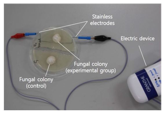
Low-voltage alternative current (AC) electrostimulation was performed using a Granthe® AC power supply (9 V, frequency: 8 Hz; Cosmic Co., Seoul, Korea) (Figure 1).
3) Statistical analysis
The areas of the colonies were determined on days 1, 4, and 7. For each level of current, the colony area of the experimental group and the control groups was analyzed using the two-way repeated-measure model. Data outcomes included the level of statistical significance within each group and within day intervals and interactions between the groups based on every interval repeatedly measured. All analyses were performed using SPSS (SPSS, 19.0 version), with p-value of <0.05 as statistically significant value.
2. Methods
All procedures were conducted using standard aseptic techniques to prevent contamination. The electrodes used in this experiment consisted of two stainless steel rods (1 mm diameter and 2.5 cm long) inserted 2.5 cm apart in the top portion of the sterile Petri plate (Figure 1). Alternate currents of 0.5 μA, 4 μA, 25 μA, 600 μA, or 900 μA were applied to assigned groups of Petri plates, starting just after inoculation with the spore suspension. Each electric stimulus was applied for 30 min at room temperature (24~26℃). The given amper-age of electric current was 0.5 μA in group A, 4 μA in B and F, 25 μA in C, 600 μA D and 900 μA in E respectively. In each plate, the fungal colony distal from the electrodes, which was at the other side of the dish, represented the control. After electric stimulation, the electrodes were removed from the plates and each Petri plate was incubated at 25℃ with 60~70% relative humidity. The effects on fungal growth were measured three times within the one-week time period: 1, 4, and 7 days after electrical stimulation. Digital photography was used to measure the colony areas of the two colonies (experimental and control) on each plate. Images were taken in a style that was reproducible, with controlled lighting, distance from the plate and background. The colony areas were calculated using Java (Image J 1.50i), which were then recalculated into ratios in order to calculate the differences among the observed colonies. The ratio signifies the current area of the colony compared to that of the previous area in order to evaluate numerical changes within the intervals.
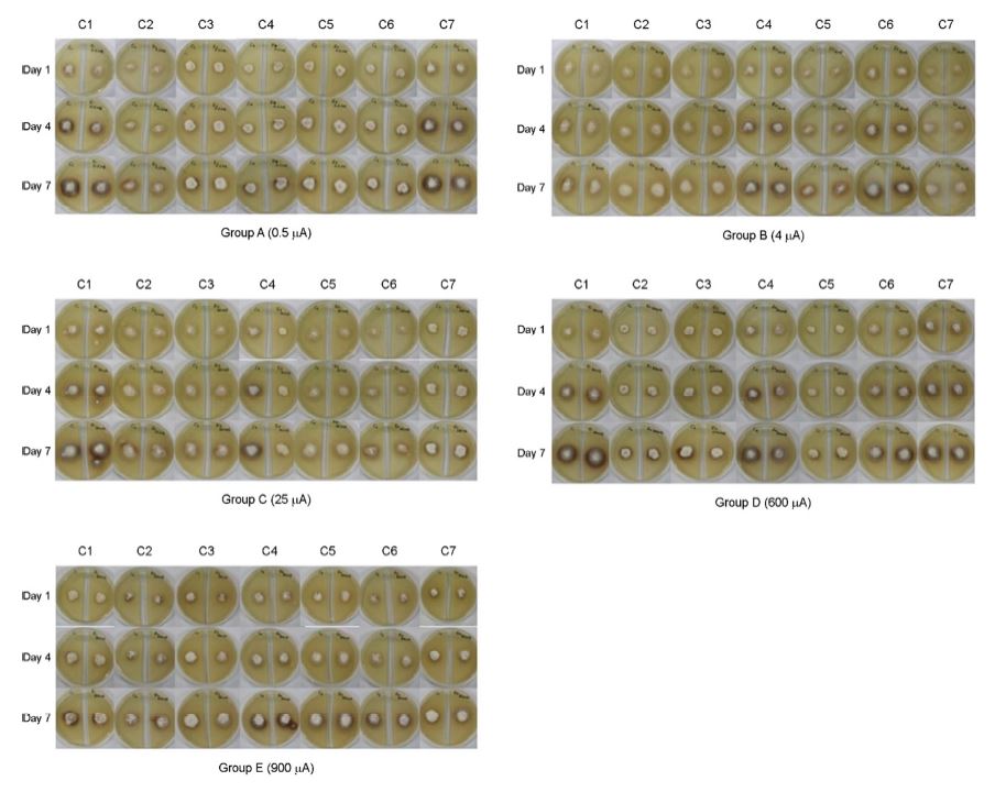
The changes in the areas of each fungal colony are depicted in Figure 2. PDACT plates were set up as two sides, with the left side representing the control group, where electrodes were not inserted, while the right side represented the experimental group where electrical stimulation was applied. There were no obvious visual differences in the sizes of the experimental fungal colonies relative to the control, regardless of the levels of current delivered (Figure 2). The colony areas at each time interval showed no statistically significant differences in colony area between the experimental and control groups (Table 1). Unlike other reported cases11,12, no evidence of gas bubble production, discoloration, temperature increase, or liquefaction around the electrodes was observed in this study.
|
Ampere |
Group |
Mean (S.D.) |
p-value (F) |
||||
|
Day 1 |
Day 4 |
Day 7 |
Time |
Group |
Time*Group |
||
|
0.5 μA |
Experimental |
0.045 (0.008) |
0.088 (0.015) |
0.131 |
<0.001 (357.767) |
0.939 (0.006) |
0.898 (0.020) |
|
Control |
0.046 (0.011) |
0.089 (0.017) |
0.131 |
||||
|
4 μA |
Experimental |
0.054 (0.005) |
0.101 (0.010) |
0.152 |
<0.001 (3023.484) |
0.788 (0.076) |
0.948 (0.008) |
|
Control |
0.053 (0.006) |
0.099 (0.010) |
0.15 |
||||
|
25 μA |
Experimental |
0.057 (0.007) |
0.107 (0.012) |
0.158 |
<0.001 (6181.368) |
0.351 (0.943) |
0.384 (0.877) |
|
Control |
0.06 (0.005) |
0.112 (0.008) |
0.163 |
||||
|
600 μA |
Experimental |
0.063 (0.011) |
0.115 (0.017) |
0.169 |
<0.001 (1515.236) |
0.960 (0.003) |
0.846 (0.045) |
|
Control |
0.064 (0.012) |
0.114 (0.018) |
0.169 |
||||
|
900 μA |
Experimental |
0.051 (0.007) |
0.098 (0.011) |
0.146 |
<0.001 (2986.533) |
0.886 (0.022) |
0.937 (0.033) |
|
Control |
0.052 (0.006) |
0.099 (0.01) |
0.147 |
||||
The growth of microorganisms depends on many factors, such as pH, osmolarity, growth medium composition, relative humidity, and temperature13,14. High concentrations of car-bohydrates and nitrogen in the medium, a pH ranging from 5 to 6, and a temperature ranging from 15℃ to 37℃, are important for fungal growth. Cornmeal agar with Tween 80 has been used to differentiate Trichophyton species from other fungal species15, while potato dextrose agar has been used to enhance the production of conidia and pigments of T. rubrum.
Microcurrent stimulation has shown various effects, including antiinflammatory effects, neuromuscular relaxation, and antibacterial effects7,9. Electricity destroys cells by disrupting the molecular structures of cell membranes, leading to irreversible changes in the membrane permeability16. Such mechanisms of membrane damage have been targeted to demonstrate antifungal effects in various in vivo and in vitro studies. However, mechanisms underlying the antifungal effect of microcurrent stimulation remain uncertain despite numerous studies being reported.
Our study sought to evaluate effects of low electric stimulation on the growth of T. rubrum and its possible medical applications in the near future. The experimental data indicated that effects of electric currents on the viability of fungi are uncertain, contrary to the previous reports. In 2004, Kalinowski et al.12 used a low-voltage direct current for up to 30 min to evaluate any inhibitory effects to fungal growth and demonstrated that clinically relevant amperages (500 μA to 3 mA) of low-voltage direct electrostimulation were fungi-static in vitro to the common pathogens of onychomycosis. In 2015, Kwon et al.10 investigated the effects of electric stimulation on fungi in a different manner. While Kalinowski et al.12 applied direct electric current to each experimental plate containing either T. rubrum or T. mentagrophytes after 24 h of incubation, Kwon et al.10 applied alternating electric current to each Petri dish immediately after the inoculation of T. rubrum, which led to faster fungal germination than the controls.
In our study, we expected the electric stimulus to modify the growth of the fungus at some level. We applied alternating currents to the inoculated plates incubated for 3 days. However, no within time interval changes were observed between the control and experimental fungal colonies (Table 1).
We failed to see any discrete increment in the growth of the inoculums throughout the experiments in both sides of the Petri dishes, indicating that the stainless steel electrode were neither fungistatic nor fungicidal.
Based on the study results, we hypothesized that the effects of electric current on fungi vary depending on the methods used to deliver the stimulation. A current delivered at a specific stage of the fungal life cycle may be as important as the intensity of the current. Contrary to the study performed by Kwon et al.10, low-voltage alternating currents may have had an effect on the fungal adherence to media rather than the rate of growth itself.
There are some limitations in this and previous studies. First, the sample sizes of the previous studies were small (12 in Kalinowski et al.12 vs. 9 in Kwon et al.10), with the studies limited to one or two fungal species (T. rubrum and T. mentagrophytes in Kalinowski et al.12 vs. T. rubrum in Kwon et al.10). Moreover, different methods were used (e.g., alternating/ direct current, different amperage, etc.), making it difficult to appropriately interpret the precise electric amperage and its antifungal effects. These factors poorly conclude the effects of electric currents on the growth of more general populations of fungi and their diversity.
In conclusion, the effects of application of electric stimulation in the form of low-voltage alternating current to fungi remain to be confirmed. A mechanism that affects the viability or growth of fungi still needs to be elucidated. As the need for alternative measures for treating fungal infections increases, further research, including in vivo studies, with larger sample sizes, diverse species, and various types of electric stimulation (direct current, alternating current, monophasic pulsed current, etc.) are imperative to clarify the possible antifungal effects of electric stimulation.
In relation to this article, I declare that there is no conflict of interest.
References
1. Surendran K, Bhat RM, Boloor R, Nandakishore B, Sukumar D. A clinical and mycological study of dermatophytic infections. Indian J Dermatol 2014;59:262-267
Crossref
Google Scholar
PubMed
2. Lee YW, Yun SJ, Lee JB, Kim SJ, Lee SC, Won YH. Clinical and mycological studies on dermatomycosis (2001-2010). Korean J Med Mycol 2013;18:30-38
Crossref
Google Scholar
3. Rodloff C, Koch D, Schaumann R. Epidemiology and anti-fungal resistance in invasive candidiasis. Eur J Med Res 2011;16:187-195
Crossref
Google Scholar
PubMed
4. Dovigo LN, Pavarina AC, Mima EG, Giampaolo ET, Vergani CE, Bagnato VS. Fungicidal effect of photodynamic therapy against fluconazole-resistant Candida albicans and Candida glabrata. Mycoses 2003;54:123-130
Crossref
Google Scholar
5. Ravanat JL, Douki T, Cadet J. Direct and indirect effects ot UV radiation on DNA and its components. J Photochem Photobiol B 2001;63:88-102
Crossref
Google Scholar
PubMed
6. Ortiz AE, Avram MM, Wanner MA. A review of lasers and light for the treatment of onychomycosis. Lasers Surg Med 2014;46:117-124
Crossref
Google Scholar
PubMed
7. Asadi MR, Torkaman G. Bacterial inhibition by electrical stimulation. Adv Wound Care (New Rochelle) 2014;3:91 -97
Crossref
PubMed
8. Kushwaha A, Shivakumar HN, Murthy SN. Iontophoresis for drug delivery into the nail apparatus: exploring hyponychium as the site of delivery. Drug Dev Ind Pharm 2016; 42:1678-1682
Crossref
Google Scholar
PubMed
9. Giladi M, Porat Y, Blatt A, Shmueli E, Wasserman Y, Kirson ED, et al. Microbial growth inhibition by alternating electric fields in mice with Pseudomonas aeruginosa lung infection. Antimicrob Agents Chemother 2010;54:3212 -3218
Crossref
10. Kwon DR, Kwon H, Lee WR, Park J. Investigating effects of nano- to micro-ampere alternating current stimulation on Trichophyton rubrum growth. Ann Dermatol 2016; 28:575-578
Crossref
Google Scholar
11. Novickij V, Grainys A, Svediene J, Markovskaja S, Paskevicius A, Novickij J. Irreversible electropermeabilization of the human pathogen Candida albicans: an in vitro experimental study. Eur Biophys J 2015;44:9-16
Crossref
Google Scholar
12. Kalinowski DP, Edsberg LE, Hewson RA, Johnson RH, Brogan MS. Low-voltage direct current as a fungicidal agent for treating onychomycosis. J Am Podiatr Med Assoc 2004;94:565-572
Crossref
Google Scholar
PubMed
13. Painter HA. Factors affecting the growth of some fungi associated with sewage purification. J Gen Microbiol 1954;10:177-190
Crossref
Google Scholar
PubMed
14. Tainwala R, Sharma YK. Pathogenesis of dermatophytes. Indian J Dermatol 2011;56:259-261
Crossref
PubMed
15. Basu S, Bose C, Ojha N, Das N, Das J, Pal M, et al. Evolution of bacterial and fungal growth media. Bioinformation 2015;11:182-184
Crossref
PubMed
16. Hülsheger H, Potel J, Niemann EG. Killing of bacteria with electric pulses of high field strength. Radiat Environ Biophys 1981;20:53-65
Crossref
Google Scholar
PubMed
Congratulatory MessageClick here!
