pISSN : 3058-423X eISSN: 3058-4302
Open Access, Peer-reviewed
pISSN : 3058-423X eISSN: 3058-4302
Open Access, Peer-reviewed

Hyun-Min Seo,Sun Gyu Kim,Yun Jung Huh,Se Uk Oh,Joung Soo Kim
10.17966/JMI.2023.28.2.23 Epub 2023 July 07
Abstract
Dermatophytosis is a fungal infection of the keratinized tissues of the skin, hair, and nails caused by dermatophytes or other fungi. Epidermophyton, Microsporum, and Trichophyton are the three genera of dermatophytes that cause skin infections. In Korea, Trichophyton rubrum is the most common dermatophytes. Dermatophytosis can be classified into anthropophilic, zoophilic, and geophilic, and most fungal infections encountered by dermatologists are tinea, dermatophytosis. This study attempted to provide a brief discussion about the clinical manifestation, diagnosis, and considerations for antifungal use in the treatment of dermatophytosis.
Keywords
Dermatophytes Dermatophytosis Superficial fungal infection Superficial mycosis Onychomycosis Tinea
Mycosis is a fungal infection caused by various fungi and can be classified into superficial, subcutaneous, deep, and systemic mycoses. Superficial mycoses refer to infections of the keratinized tissues of the skin, hair, and nails caused by dermatophytes; however, infections caused by fungi other than dermatophytes are also included in this definition. Dermatophytes that cause skin infections are classified into three genera: Epidermophyton, Microsporum, and Tricho- phyton1. Trichophyton rubrum, Trichophyton mentagrophytes, Trichophyton verrucosum, and Trichophyton tonsurans are the most common dermatophytes in Korea, and Microsporum canis was reported in small numbers2. Candida or Malassezia infections that cause superficial fungal infections are separately defined as superficial yeast infections. Fungi can be classified taxonomically into anthropophilic, zoophilic, and geophilic. Anthropophilic fungi are characterized by the absence or presence of mild inflammation because they have adapted to humans as hosts for a long time. Zoophilic and geophilic fungi are characterized by severe inflammation because they do not primarily infect humans as hosts. Among zoophilic fungi in Korea, M. canis is a representative fungus that mainly infects dogs and cats3. As a dermatologist, most of the fungal infections encountered by dermatologists are superficial mycoses. Therefore, this manuscript will cover superficial mycoses, including nail infections.
Tinea capitis
Tinea capitis is typically more common in children aged 3~14 years, and its incidence decreases during adolescence when sebum secretion increases. This is thought to be caused by fatty acids in the sebum that have bacteriostatic activity against fungi. In Korea, the incidence of tinea capitis has decreased over time; however, some patients in whom infection developed after menopause may still visit hospitals because of hair loss. The most reported causative fungi in cases of tinea capitis in Korea is M. canis, which often causes severe inflammation2,3. The second most common causative fungi is T. rubrum, which is characterized by the presence of numerous scalp scales. T. tonsurans, which forms arthro- coninia within the hair shaft (endothrix), is the predominant species in the United States, and it often presents as a pattern of clustered, broken hairs4. On the contrary, M. canis and T. rubrum often do not exhibit broken hairs as the main form because they form arthroconinia outside the hair (ectothrix)5.
If severe inflammation of the scalp is present, permanent scarring of the scalp may remain after treatment6. Therefore, tinea capitis should be orally treated with antifungal agents, and depending on the severity of inflammation, systemic steroids can be appropriately added. Even if alopecia occurs immediately after treatment, some degree of recovery can be expected, except in very severe areas7. Thus, the potassium hydroxide (KOH) test, as a diagnostic method, should be performed8. Fungus culture can identify the causative fungi. In cases caused by M. canis, yellow-green fluorescence can be observed with Wood's lamp.
Superficial mycoses of the skin other than the scalp
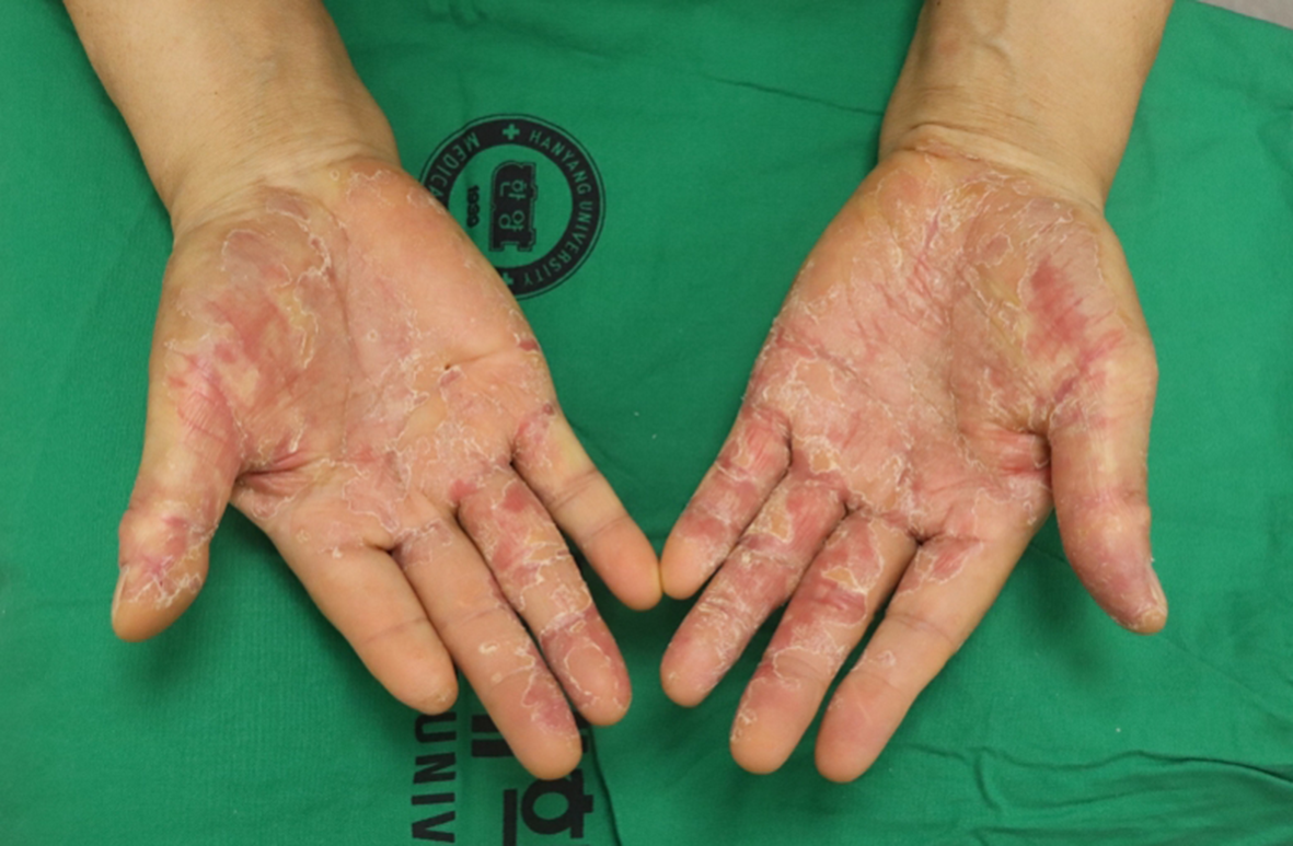
Tinea barbae is a specific term used when superficial mycosis occurs in bearded areas. Tinea barbae occurring in sites other than bearded areas, such as the face, trunk, groin, and hand/feet, is called tinea faciale, tinea corporis, tineacruris, and tinea manuus/pedis, respectively9 (Fig. 1). While differences in characteristics depending on the body part, the diagnosis and treatment are generally similar. Typically, the clinical manifestation involves lesions with abundant scales and without significant inflammation. They often appear as circular or annular rash that spreads peripherally and can be diagnosed through the KOH test. Superficial mycoses in closed areas require caution because they may be accompanied by secondary infection. Severe dermatitis accompanied by the primary lesion can often cause an id reaction, resulting in the spread of the rash to other body areas. If a rash spreads to other body parts, patients who may be concerned about systemic fungal infections must be reassured, as it does not involve any fungal hyphae or spores.
Tinea unguium
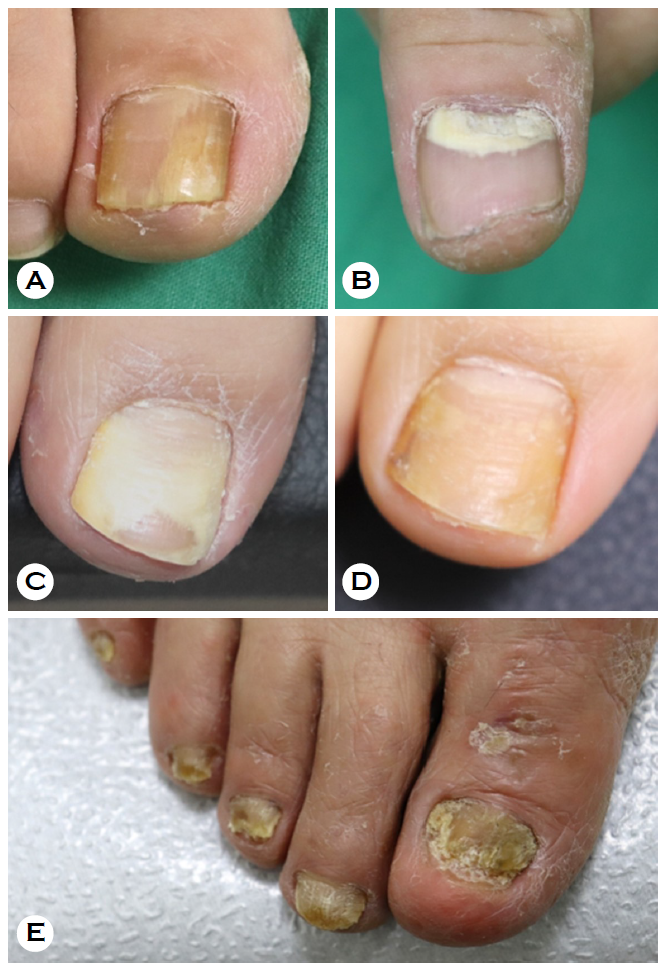
Tinea unguium is a nail infection caused by dermatophytes, and onychomycosis is used to describe all fungal infections of the nails, including those caused by non-dermatophyte fungi. In most cases, it is caused by dermatophyte infections; thus, the two terms are often used interchangeably. Onychomycosis is classified as distal-lateral subungual, proximal subungual, white superficial, and total dystrophic onychomycoses (Fig. 2A-2E)10. The distal-lateral subungual type is the most com- mon form of onychomycosis, while the proximal subungual type is rare and indicate the need for screening for human immunodeficiency virus infection11. The diagnosis of onychomycosis involves KOH tests and fungus culture on the sus- pected lesion. Additionally, diagnostic tests such as KOH-treated nail clipping stained with periodic acid-Schiff (KONCPA) and tinea unguium rapid antigen test can also be helpful in the diagnosis12,13.
TREATMENT OF SUPERFICIAL MYCOSES
Superficial mycoses, excluding tinea capitis and onycho- mycosis, are usually easy to treat, and management focuses more on preventing recurrence than on treatment. As fungi generally thrive in humid environments, the infected area must be kept dry. In cases of excessive sweating, aluminum chloride solutions may be used to regulate perspiration. When applying topical antifungal agents, it is recommended to apply the medication to the surrounding area within 6 centimeters, twice a day. The treatment period ranges from 2 weeks to 6 weeks until the suspected lesion is completely resolved, and as the duration of treatment increases, the success rate also increases14. For foot infections, patients should be educated to apply the medication thoroughly, including areas between the toes. To prevent reinfection from contaminated dead scales, socks should be turned inside out when laundering. Soaking clothes in a diluted solution of water and bleach can also be effective; however, care should be taken to avoid damaging the fabric with the bleach. Topical imidazole antifungal agents are commonly used for treating superficial mycoses. Amorolfine, a morpholine anti- fungal agent, is also widely used. Failed treatments are often related to external factors such as incorrect application or reinfection rather than inadequate drug efficacy. To increase the treatment success rates and prevent a recurrence, the use of an antifungal shampoo is also effective when washing the infected area, in addition to applying topical antifungal agents15.
Tinea capitis and moderate-to-severe onychomycosis should be treated with oral antifungal therapy, and in dermatophyte infections, terbinafine was reported to have higher success rates than azole drugs16. Drug-induced liver injury (DILI) is a well-known adverse reaction to oral antifungal agents. In a large cohort study of 69,830 patients treated with oral antifungal agents, DILI occurs in 134.1 per 100,000 person-months for ketoconazole, 10.4 for itraconazole, and 2.5 for terbinafine, presenting that terbinafine has the lowest risk of DILI17. Itraconazole is well known for its contraindication with statins (HMG-CoA reductase inhibitors), which are com- monly used as cholesterol-lowering drugs. Therefore, it is often difficult to use in middle-aged and older age groups, and it cannot be used in patients with congestive heart failure and ventricular dysfunction18,19. Although no absolute contraindications were established for terbinafine in terms of drug interactions, the dose should be halved in patients with acute or chronic kidney diseases with a creatinine clearance of ≤50 mL/min20. Based on these considerations, terbinafine is the first-line choice because of its excellent efficacy, min- imal drug interactions, and low frequency of adverse drug reactions. Itraconazole and fluconazole are the second- and third-line drugs, respectively16. However, in patients with superficial mycoses other than dermatophytes, itraconazole and fluconazole, which are broad-spectrum drugs, are pre- ferred. Recently developed drugs, such as oteseconazole and fosravuconazole, have shown good efficacy in patients with onychomycosis21,22. However, a previous study reported that fosravuconazole can cause mild elevation of liver enzymes; thus, further large-scale studies are needed22.
The use of topical agents is an important treatment option for patients with onychomycosis who cannot take antifungal agents orally. When applied topically for 48 weeks, efinaconazole has demonstrated good efficacy and similar therapeutic effects as taking itraconazole orally for 12 weeks23. Other options such as ciclopirox and amorolfine nail lacquer are also available in Korea. Physical treatments such as the application of urea ointment or nail avulsion may be con- sidered when topical agents are not effective. In a recent meta-analysis, combination therapy of topical antifungal agents and laser therapy was reported to be more effective than topical antifungal agents alone24.
This brief review focuses on the clinical manifestation, diagnosis, and treatment of superficial mycoses, which have a high prevalence and commonly relapse. Therefore, it is crucial to not only focus on the diagnosis and treatment but also on preventing recurrence.
References
1. Leung AKC, Lam JM, Leong KF, Hon KL, Barankin B, Leung AAM, et al. Onychomycosis: An updated review. Recent Pat Inflamm Allergy Drug Discov 2020;14:32-45
Google Scholar
2. Lee WJ, Eun DH, Jang YH, Bang YJ, Jun JB. The incidences of dermatophytosis and cutaneous candidiasis infection in Southeastern Korea between 2013 and 2016. J Mycol Infect 2018;23:1-8
Google Scholar
3. Cheon SJ, Lee JH, Lee YW, Park J, Suh MK, Kim H, et al. Epidemiology and identification of organisms causing superficial dermatomycoses at tertiary hospitals in Korea: A prospective multicenter study. J Mycol Infect 2018;23: 45-53
Google Scholar
4. Patel GA, Schwartz RA. Tinea capitis: still an unsolved problem? Mycoses 2011;54:183-188
Google Scholar
5. Zhu M, Li L, Wang J, Zhang C, Kang K, Zhang Q. Tinea capitis in Southeastern China: a 16-year survey. Myco- pathologia 2010;169:235-239
Google Scholar
6. Mirmirani P, Willey A, Chamlin S, Frieden IJ, Price VH. Tinea capitis mimicking cicatricial alopecia: what host and dermatophyte factors lead to this unusual clinical presentation? J Am Acad Dermatol 2009;60:490-495
Google Scholar
7. Seo HM, Park SK, Park JH, Kim KY, Kim JS. Scarring alopecia after kerion celsi in adults. J Mycol Infect 2022: 21-22
Google Scholar
8. Park J, Nam JH, Lee JH, Park J, Mun JH, Lee YW, et al. Korean guideline for the diagnosis and treatment of onychomycosis: Purpose and process of algorithm guide- line development. J Mycol Infect 2018;23:33-44
Google Scholar
9. Drake LA, Dinehart SM, Farmer ER, Goltz RW, Graham GF, Hardinsky MK, et al. Guidelines of care for superficial mycotic infections of the skin: tinea corporis, tinea cruris, tinea faciei, tinea manuum, and tinea pedis. Guidelines/ Outcomes Committee. American Academy of Derma- tology. J Am Acad Dermatol 1996;34:282-286
Google Scholar
10. Hay RJ, Baran R. Onychomycosis: a proposed revision of the clinical classification. J Am Acad Dermatol 2011;65: 1219-1227
Google Scholar
11. Prose NS, Abson KG, Scher RK. Disorders of the nails and hair associated with human immunodeficiency virus infection. Int J Dermatol 1992;31:453-457
Google Scholar
12. Haghani I, Shokohi T, Hajheidari Z, Khalilian A, Aghili SR. Comparison of diagnostic methods in the evaluation of onychomycosis. Mycopathologia 2013;175:315-321
Google Scholar
13. Paugam A, Challier S. Dermatophytic onychia: Effective- ness of rapid immunochromatographic diagnostic testing directly on samples compared to culture. Ann Dermatol Venereol 2022;149:108-111
Google Scholar
14. Crawford F, Hollis S. Topical treatments for fungal in- fections of the skin and nails of the foot. Cochrane Data- base Syst Rev 2007;2007:CD001434
Google Scholar
15. Ward H, Parkes N, Smith C, Kluzek S, Pearson R. Con- sensus for the treatment of tinea pedis: A systematic review of randomised controlled trials. J Fungi (Basel) 2022;8:351
Google Scholar
16. Fávero MLD, Bonetti AF, Domingos EL, Tonin FS, Pontarolo R. Oral antifungal therapies for toenail onycho- mycosis: a systematic review with network meta-analysis toenail mycosis: network meta-analysis. J Dermatolog Treat 2022;33:121-130
17. García Rodríguez LA, Duque A, Castellsague J, Pérez-Gutthann S, Stricker BH. A cohort study on the risk of acute liver injury among users of ketoconazole and other antifungal drugs. Br J Clin Pharmacol 1999;48:847-852
Google Scholar
18. de Sá DC, Lamas AP, Tosti A. Oral therapy for onycho- mycosis: an evidence-based review. Am J Clin Dermatol 2014;15:17-36
Google Scholar
19. Lipner SR, Scher RK. Onychomycosis: Treatment and pre- vention of recurrence. J Am Acad Dermatol 2019;80: 853-867
Google Scholar
20. Gupta AK, Venkataraman M, Talukder M. Onychomycosis in older adults: Prevalence, diagnosis, and management. Drugs Aging 2022;39:191-198
Google Scholar
21. Elewski B, Brand S, Degenhardt T, Curelop S, Pollak R, Schotzinger R, et al. A phase II, randomized, double-blind, placebo-controlled, dose-ranging study to evaluate the efficacy and safety of VT-1161 oral tablets in the treatment of patients with distal and lateral subungual onychomycosis of the toenail. Br J Dermatol 2021;184: 270-280
Google Scholar
22. Watanabe S, Tsubouchi I, Okubo A. Efficacy and safety of fosravuconazole L-lysine ethanolate, a novel oral triazole antifungal agent, for the treatment of onycho- mycosis: A multicenter, double-blind, randomized phase III study. J Dermatol 2018;45:1151-1159
Google Scholar
23. Elewski BE, Rich P, Pollak R, Pariser DM, Watanabe S, Senda H, et al. Efinaconazole 10% solution in the treat- ment of toenail onychomycosis: Two phase III multicenter, randomized, double-blind studies. J Am Acad Dermatol 2013;68:600-608
Google Scholar
24. Zhang J, Lin P, Li J, Guo C, Zhai J, Zhang Y. Efficacy of laser therapy combined with topical antifungal agents for onychomycosis: a systematic review and meta-analysis of randomised controlled trials. Lasers Med Sci 2022;37: 2557-2569
Google Scholar
Congratulatory MessageClick here!
